
Tuhin Mistry1, Rajni Mathur2, Narender Saini3, Pratibha Rathore4
Tuhin Mistry1, Rajni Mathur2, Narender Saini3, Pratibha Rathore4
1 Junior Resident; 2Professor; 4Assistant Professor
Department of Anesthesiology and Critical Care
3Assistant Professor; Department of Orthopedic Surgery
Sawai Man Singh Medical College and Attached Group of Hospitals, Jaipur, Rajasthan (India)
Correspondence:
Dr. Tuhin Mistry, Room No. A-92, RD Hostel, SMS Medical College and Hospital, Jaipur, Rajasthan (India); E-mail: dr.tuhin2014@gmail.com
ABSTRACT
Amniotic band syndrome (ABS) is a rare condition, potentially associated with a variety of different birth defects. It is also known as ADAM complex (amniotic deformities, adhesion, mutilation), amniotic band sequence, amniotic disruption complex, annular grooves, congenital amputation, congenital constricting bands, Streeter bands, Streeter anomaly, transverse terminal defects of limb, aberrant tissue bands, amniochorionic mesoblastic fibrous strings, and amniotic bands.1 The severity of amniotic band syndrome can range from a single, isolated finding to multiple, disfiguring complications. The arms and legs are most often affected. The head and face and, in some cases, various internal organs can also be affected. General anesthesia is the preferred option for surgical corrections of the anomalies.
Key words: Amniotic band syndrome; birth defects; Clift lip; Cleft palate
Citation:
Mistry T, Mathur R, Saini N, Rathore P. Perioperative management of amniotic band syndrome: a case report and literature review. Anaesth Pain & Intensive Care 2015;19(4):505-509
INTRODUCTION
Amniotic band syndrome (ABS) is a set of congenital malformations ranging from minor constriction rings and lymphedema of the digits to complex, bizarre multiple congenital anomalies that have been attributed to amniotic bands that stick, entangle and disrupt fetal parts. The estimated incidence of ABS ranges from 1:1200 to 1:15,000 in live births2-4 and 1:70 in stillbirths.5 It affects both sexes equally. The anesthetic management is usually straight forward and general anesthesia is the choice. We present this case of ABS mainly because of academic interest.
CASE REPORT
A male baby of 7 months and 8 kg body weight was admitted for treatment of constriction rings affecting all of his four limbs. It was also associated with cleft lip and bilateral cleft palate. He had been operated earlier at 4 months of age for cleft lip and was awaiting correction of cleft palate. He was a full-term baby, born normally at hospital to a G3 P2, 30 year-old mother. There was no significant antenatal history. First antenatal ultrasonography was done in the third month of pregnancy, which was normal. His developmental milestones were within normal limits. He had difficulty in feeding and nasal regurgitation due to cleft palate, but no history of pulmonary or ear infection. Clinical examination revealed a constriction mark over right and left arm 2 cm proximal to wrist joint, right and left lower limbs 3 cm above the ankle joint in total circumference and associated congenital talipes equinovarus (TEV) on left side, bilateral cleft palate and corrected cleft lip (Figures 1-6).
His axillary, brachial, radial, ulnar, femoral, popliteal and dorsalis pedis arterial pulsations were normal on both sides. All the fingers and toes were normal. Motor and sensory functions of all four limbs were
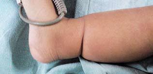 Figure 1: A partial band on right leg (medial side)
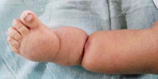 Figure 2: A complete band on left leg
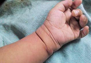 Figure 3: A band on left fore arm
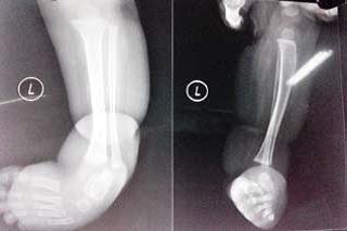 Figure 4: Left leg; AP and lateral views
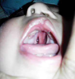 Figure 5: Associated cleft lip & palate with nasal deformity
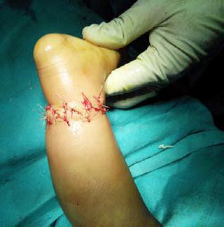
Figure 6: Z-plasty done
also normal. Other body systems were normal. Routine laboratory investigations, e.g. hemoglobin, hematocrit, white blood cell count, electrolytes and urine analysis were within normal limits. Chest x-ray (PA view), 12 leads ECG, USG whole abdomen and 2D echocardiography were all within normal limits. Skiagram of his limbs showed soft tissue constriction without involvement of the bones. Doppler study of the limbs revealed normal blood flow in all the vessels, as well as at the constriction site. Most severe constrictions were present on both legs and he was operated for that by excising the constriction band and multiple Z-plasty under general anesthesia.
Preoperative airway assessment revealed adequate mouth opening with bilateral cleft palate, Mallampati grade II. The thyromental distance, sternomental distance, atlanto-occipital joint and the TM joint movements were within normal limits. On the day of operation he was in fasting state for 4 hours. In the OT, standard monitors including pediatric pulse oximeter, NIBP cuff, 5-lead ECG were attached. Preoperatively his pulse rate was 121 beats / min, NIBP 90/60 mmHg and O2 saturation was 100% on room air. An intravenous line was secured with 24 gauge cannula and inj. ringer lactate started via pediatric drip set. He was preoxygenated with 100 % oxygen via facemask for 10 min. Premedication consisted of inj. midazolam 0.03 mg/kg, glycopyrrolate 0.01 mg/ kg and fentanyl 2 μg/kg. Induction was performed with 3 mg/kg of thiopentone sodium and 2 mg/kg succinylcholine. Orotracheal intubation with a 4.0 mm internal diameter (ID) uncuffed endotracheal tube was performed under direct laryngoscopy (Cormac and Lehane Grade III, Grade II intubation). Position of ET tube was confirmed by 5 point auscultation and EtCO2. The tube was fixed at right angle of mouth at 12 cm mark with adhesive tapes. The patient was on controlled ventilation. Anesthesia was maintained with N2O:O2 (50:50), isoflurane and atracurium 0.1 mg/kg. Antibiotic was given before skin incision. To prevent hypothermia, his extremities were adequately covered with warm blankets, room temperature was increased and warm IV fluids were used. Surgery was continued for one hour. After the surgery neuromuscular blockade was reversed with inj. neostigmine 0.08 mg/kg plus inj. glycopyrrolate 0.01 mg/kg. Tracheal extubation was done. The postoperative period was uneventful and he was discharged after 3 days.
DISCUSSION
Amniotic constriction bands are strands of the amniotic sac that surrounds a baby in the womb. They may cause a congenital deformity of the face, arms, legs, fingers, or toes. Although there are many unanswered queries regarding congenital constriction band syndrome, studies regarding its cause have came a long way. There are two main theories.6-9 Widely accepted “extrinsic model” theory, proposed by Torpin et al10 in 1968 explains, the rupture of the amnion without the rupture of the chorion leads to transient oligohydramnios due to loss of amniotic fluid through the initially permeable chorion. The fetus passes from the amniotic to the extra embryonic coelom through the defect and comes in contact with ‘sticky’ mesoderm on the chorionic surface of the amnion. This leads to entanglement of the fetal parts and skin abrasions. Entanglement of the fetal parts causes constriction rings and amputations, whereas skin abrasions can lead to disruption defects, such as cephaloceles. Further, swallowing of the bands will cause asymmetric clefts on the face. The “intrinsic model” was proposed by Streeter in 1930 and suggests that the anomalies and the fibrous bands have a common origin, caused by a perturbation of developing germinal disc of the early embryo.
Most cases of ABS are not of genetic origin, and occur sporadically with no recurrence in siblings or children of affected adults.11 Maternal trauma, oophorectomy during pregnancy,12 intrauterine contraceptive device13 and amniocentesis14 and familial incidence of connective tissue disorders (Ehler-Danlos syndrome)15 are some of the implicated etiopathological factors. Lockwood C et al suggested the role of a teratogenic insult as one of the factors causing the disorder.16
Amniotic band syndrome has very polymorphic clinical findings. Early amniotic rupture, during first 45 days, leads to the most severe cranio-facial and visceral malformations.17 The most common finding in the amniotic band syndrome is the constriction rings of the fingers and toes.18 It occurs in 77 percent cases with multiple anomalies.19 In our case, there were constriction rings of the soft tissues accompanied by distal edema. Other associated features of ABS are shortening of the limbs or intrauterine limb amputation, amputation of the digits (most often 2nd, 3rd and 4th fingers) and toes, syndactyly, hypoplasia of the digits, foot deformities, pseudarthrosis, peripheral nerve palsy.2,20 If bands compress the fetal head or face, different cranio-facial disturbances appear like asymmetric face clefts, orbital defects (anophthalmos, microphthalmos, enophthalmos), corneal abnormalities, central nervous system malformations (anencephaly, encephalocele, asymmetric meningocele) and calvaria defect etc. Amniotic bands can also cause abdominal wall defect and abdominal organs extrophy,2 chest wall defect with heart extrophy,6 umbilical cord strangulation with often lethal outcome.2 Amniotic rupture and consecutive oligoamnios can cause deformities such as metatarsovarus, scoliosis5 or hip dislocation2 by mechanical pressure on the fetus. Because of such a wide spectrum of possible anomalies and many combinations of their simultaneous appearance, there are no two identical cases of ABS.2 Beside all previously mentioned malformations caused by amniotic bands itself, a subset of cases manifest additional findings that are not consistent with that mechanism, such as congenital heart defects, renal anomalies, hemangiomas, imperforate anus, polydactyly, septo-optic dysplasia, typical cleft lip and palate. 6 The association of abnormalities, such as club feet, anencephaly, cleft lip, cleft palate, rib clefting, gastroschisis, omphalocele, bladder exstrophy and imperforate anus should be regarded as strong evidence and should arouse the suspicion of ABS. In our case, the child had had associated cleft palate and cleft lip.
ABS is often difficult to detect before birth, although the earliest may be difficult to detect by ultrasound, and are more often diagnosed by the effects they have on fetal anatomy. The visualization of amniotic bands attaching to a fetus with restriction of motion, constriction rings on extremities and irregular amputations of fingers and/or toes with terminal syndactyly on prenatal ultrasonography is diagnostic of the condition. The visualization of amniotic bands or sheets in the absence of fetal deformities should by no means lead to the diagnosis of the ABS since several types of membranes such as folds of amnion may be seen in normal pregnancies. 21 Latest ultrasound techniques – 3D and 4D ultrasound contribute to more sensitive prenatal diagnostics of ABS, and in complicated cases fetal MRI can be helpful.3 Doppler study of the constricted limb could be of useful in diagnosis of in-utero amputation, and could be helpful to determine when inutero treatment should be considered. Physical examination is the main way of postnatal diagnosis of ABS, with a search to establish potential malformations of different organs and body parts. Ultrasound, echocardiography, and x-ray films may help diagnose or rule out other associated anomalies.
Management strategy of ABS depends upon the extent of the associated anomalies. Treatment is mostly surgical with an individual approach to every single case. Termination of pregnancy is usually proposed at the time of the diagnosis of severe craniofacial and visceral abnormalities (fetal anomalies incompatible with life), whereas minor limb defects can be repaired with postnatal surgery. 22 Lately, there have been some attempts of prenatal ABS treatment – fetoscopic laser cutting of amniotic bands, before their compression on the fetus makes malformations.23 Patterson24 in his study of 52 patients of congenital constriction rings had reported only 2 below knee amputations in addition to other musculoskeletal defects. Zych, et al25 in 1983 reported a case of involvement of congenital bands, pseudarthrosis and impending gangrene of leg, which was salvaged with multiple Z-plasty. Greene WB26 in his study advised a one-stage release for circumferential congenital constriction bands which was performed in four extremities. Samra et al27 in 2006 reported a case of severe constricting amniotic band with a threatened lower extremity in a neonate, which was salvaged with multiple Z-plasties after a 6-year functional follow up. So, the outcome of the disease depends on the gravity of the malformation.
Our case was operated in single stage, the one-stage release facilitated postoperative care and there was no need for additional periods of anesthesia or for additional operations, which are necessary when this problem is treated in two or three stages. Amniotic constriction band presents at birth should be treated as early as possible, because it becomes more pronounced as time passes. Recently, constriction ring has successfully been released by a minimally invasive endoscopic surgical technique avoiding severe limb dysfunction or foot amputation.28 Congenital band may cause edema of the tissue distal to the band, they are distinguished from congenital lymph edema. If edema persists after surgical correction of the bands, excision of the edematous area may be necessary with direct closure or conversion of the overlying skin to thick and free by partial thickness skin grafts. Our case presented at an age of 7 months and there were no threatened complications like gangrenous changes, bony deformity or loss of sensation or function and it was treated in single stage by multiple Z-plasty.
Recently, for postnatal surgical correction of constriction rings, a new technique (Mutaf procedure) has been reported, which advices filling the circular groove caused by the constriction ring with dermofat flaps. This eliminates the soft tissue deficiency and provides a normal extremity contour. Moreover, since rectangular-plasty allows replacing the major limbs of the incisional scars within the relaxed skin tension lines, it provides a better scar in comparison with old Z-plasty techniques.29
Regarding anesthetic management, child should be evaluated properly for any hidden systemic anomaly during pre- anesthetic check-up. Anesthetist should always be prepared for difficult airway in such cases. In addition to deformities in ABS, pediatric airway is challenging because of unique anatomy (smaller size, vocal cord between C1-C4 with an anterior angulation, a large and floppy epiglottis, a large occiput) and physiology (frequent upper airway obstruction under GA, higher metabolism, and faster desaturation during period of apnea). In our case, it was a difficult intubation as it required external manipulation. Difficult airway cart with different sizes of endotracheal tubes, laryngeal mask airway, pediatric fiberscope etc. were kept ready although it was not required. To prevent hypothermia we took several measure like adequate covering of extremities with warm blankets, elevation of room temperature and warm IV fluids. Extubation should be done when there is regular spontaneous breathing, vigorous movements of all limbs, good oxygen saturation and absence of significant hypothermia.
CONCLUSION
ABS is rare seen in routine practice, but in every newborn with congenital anomalies, defect of extremities and/or body walls must be looked for. Because of its complexity, the treatment and follow-up of these neonates requires a team of specialists depending upon the special needs of each patient.
REFERENCES
- Luis Flavio Gonçalves, Philippe Jeanty. Amniotic band syndrome. 1999-09-26-18
- Poeuf B, Samson P, Magalon G. Amniotic band syndrome. Chir Main 2008 Dec;27 Suppl 1:S136-47.
[PubMed] doi: 10.1016/j.main.2008.07.016.
- Merrimen JL, McNeely PD, Bendor-Samuel RL, Schmidt MH, Fraser RB. Congenital placental–cerebral adhesion: an unusual case of amniotic band sequence. J Neurosurg 2006;104(5 Suppl): 352–5
- Jabor MA, Cronin ED. Bilateral cleft lip and palate and limb deformities: a presentation of amniotic band sequence? J Craniofac Surg 2000;11(4):388-93
- Ross MG. Pathogenesis of amniotic band syndrome. Am J Obstet Gynecol.2007;197(2):219-20
- Robin NH, Franklin J, Prucka S, Ryan AB, Grant JH. Clefting, amniotic bands and polydactyly: A distinct phenotype that supports an intrinsic mechanism for amniotic band sequence. Am J Med Genet A. 2005;137A(3):298-301.
- Lateo SA, Taylor AE, Meggitt SJ. Raised limb bands developing in infancy. Br J Dermatol 2006;154(4):791-2. [PubMed]
- Werler MM, Louik C, Mitchell AA. Epidemiologic analysis of maternal factors and amniotic band defects. Birth Defects Res A Clin Mol Teratol 2003;67(1):68-72.
- Fathallah ZF. Unusual Presentation of Amniotic Band Syndrome. Bas J Surg 2007;11:77-9
- Torpin R. Fetal malformations caused by amnion rupture during gestation. Springfield (IL): Charles C Thomas; 1968. p. 1–76.
- Verma A, Mohan S, Kumar S. Late presentation of amniotic band syndrome: A case report. J Clin Diag Res. 2007;1:65-68.
- Tanaka O, Koh T, Otani H. Amniogenic band anomalies in a fifth-month fetus and in a newborn from maternal oophorectomy during early pregnancy. Teratology 1986;33:187–93.
- Csecsek K, Szeifert GT, Papp Z. Amniotic bands associated with early rupture of amnion due to an intrauterine device. Zentralbl Gynakol 1987;109:378–41.
- Kohn G. The amniotic band syndrome: a possible complication of amniocentesis. Prenat Diagn 1987;8:303–5. [PubMed]
- Young ID, Lindenbaum RH, Thompson EM, Pembrey ME. Amniotic band syndrome in connective tissue disorders. Arch Dis Child 1985;60:1061–3.
- Lockwood C, Ghidini A, Romero R, Hobbins JC. Amniotic band syndrome: reevaluation of its pathogenesis. Am J Obstet Gynecol 1989;160:1030–3.
- Jabor MA, Cronin ED. Bilateral cleft lip and palate and limb deformities: a presentation of amniotic band sequence? J Craniofac Surg 2000;11(4):388-93
- Fiedler JM, Phelan JP. The amniotic band syndrome in monozygotic twins. Am J Obstet Gynecol 1983;14:863–4.
- Nyberg DA, Mahony BS, Pretorius DH. Diagnostic ultrasound of fetal anomalies. Littleton (MA): Year Book Medical Publishers; 1990.
- McGuirk CK, Westgate MN, Holmes LB. Limb Deficiencies in Newborn Infants. Pediatrics 2001;108(4):e64-e70.
- Pregnancy: a trip to the extraembryonic coelom. J Ultrasound Med 1990;9:733–6.
- Sentilhes L, Verspyck E, Patrier S, Eurin D, Lechevallier J, Marpeau L. Amniotic band syndrome: pathogenesis, prenatal diagnosis and neonatal management. J Gynecol Obstet Biol Reprod (Paris) 2003;32:693-704.
- Quintero RA, Morales WJ, Phillips J, Kalter CS, Angel JL. In utero lysis of amniotic bands. Ultrasound Obstet Gynecol 1997;10(5):316-20.
- Patterson TJ. Congenital ring-constrictions.Br J Plast Surg 1961;14:1-31
- Zych GA, Ballard A. Constriction band causing pseudarthrosis and impending gangrene of the leg. A case report with successful treatment. J Bone Joint Surg 1983;65A:410-412
- Greene WB. One stage release of congenital constriction bands. J Bone Joint Surg Am 1993;75:650-665 [PubMed]
- Samra S, Samra AH. Threatened lower extremity in a neonate from a severely constricting amniotic band. Ann Plast Surg 2006;57:569-572
- Ronderos-Dumit D, Briceño F, Navarro H, Sanchez N. Endoscopic release of limb constriction rings in utero. Fetal Diagn Ther 2006; 21:255-258
- Mutaf M, Sunay M. A new technique for correction of congenital constriction rings. Ann Plast Surg 2006;57:646 – 52.
