
Kalpana Rajendra Kulkarni, MBBS, DNB, DA*, Basant Rambharose Chaurasia, MBBS, MD**
*Professor of Anesthesiology; **Senior Resident
Department of Anesthesiology, D Y Patil Medical College & Hospital, Kadamwadi, Kolhapur, Maharashtra (India)
Correspondence: Dr.Mrs.Kalpana R.Kulkarni, 1168, A-5, “Chaitanya”, Takala Square, Kolhapur- 416008. (Maharashtra) INDIA; Mobile: 9822065665; E-mail: drrmk@rediffmail.com
ABSTRACT
Retropharyngeal abscess occurs most commonly in children following acute upper respiratory tract or ear infection. We report a case of difficult airway due to retropharyngeal abscess in five years old female child, posted for incision and drainage under general anesthesia. Following inhalational induction with 6% sevoflurane in 100% oxygen, intubated with 4.5 mm ID plain Portex® endotracheal tube.
Anesthesia was maintained with 50% O2 in N2O and rocuronium 10 mg with traces of sevoflurane. Intraoral and lateral neck incisions were taken to drain the abscesses and extubated following satisfactory clinical recovery. However immediately after extubation the child developed severe stridor and airway obstruction leading to desaturation down to 60% of SpO2. Three attempts for reintubation under the effect of 12.5 mg intravenous succinylcholine failed. Hence airway secured with emergency tracheostomy. Here we discuss about the anesthetic management and the immediate post operative crisis due to difficult reintubation.
Keywords: Retropharyngeal abscess; Difficult airway; Tracheostomy
Citation: Kulkarni KR, Chaurasia BR. Difficult airway in a child with retropharyngeal abscess. Anaesth Pain & Intensive Care 2014;18(3):285-88
INTRODUCTION
Retropharyngeal abscess (RPA) develops in children of < 6 years of age due to suppuration of retropharyngeal lymph nodes following upper respiratory tract /ear infection, trauma or foreign body. The symptoms are sore throat, fever, neck stiffness and stridor. It needs aggressive medical management and/or surgical drainage under general anesthesia which is associated with higher incidence of morbidity and mortality.1 We report a child with RPA, who developed post extubation stridor and desaturation . Emergency tracheostomy was performed to manage ‘can’t intubate – can’t ventilate’ scenario.
CASE REPORT
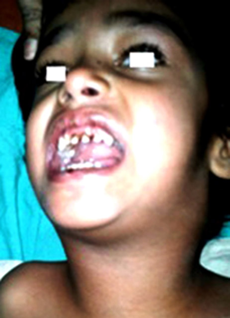
A 12 Kg, 5 years old female child, had cough with fever, swelling in the neck region and difficulty in swallowing since 5 days. She had similar episode 15 days back with macular rash and thrombocytopenia, which responded to the medication. Again she got admitted in PICU for dyspnea with acute stridor, desaturation (SpO2 of 85%), needed intubation and ventilatory support on SIMV mode for 72 hours and was extubated following response to the antibiotics. Later within 2 days, her neck swelling increased in size and on investigations the diagnosis of RPA was confirmed, hence posted for drainage under GA. There was no history of trauma or ingestion of foreign body (Figure 1).
Herblood analysis showed Hb 8.1 gm/dl and TLC raised to 22400/mm.3 Coagulation profile was within normal limits, however, she had a lower sodium level and lower albumen. Chest x-ray PA view and x-ray neck AP/lateral views on admission revealed no abnormality. On 5th day an USG neck reported multiple cervical enlarged lymph nodes, panadenitis. CT scan revealed multiple fluid level density lesion in retropharyngeal region, communicating abscesses in bilateral parapharyngeal, carotid and parotid spaces extending from base of skull up to the larynx. Enlarged necrotic level II, III in posterior triangle group of lymph nodes. No foreign body was detected(Figure 2).
On general examination she was irritable, febrile, bilateral neck swelling with restricted neck mobility. Mouth opening was 2.5cm with grade III Mallampati score. Her heart rate (HR) was 154/min, respiratory rate (RR) of 26/min and oxygen saturation (SpO2) of 99% on room air. On Indirect laryngoscopy there was edematous epiglottis, aryepiglottic fold, pink narrow glottic opening with visible false vocal cords. On systemic examination no abnormality detected.
Figure 1: Child with retropharyngeal abscesses
Anesthetic Management: Following informed high-risk consent and six hours of fasting, vital sign monitoring was started with SpO2, ECG and NIBP. Difficult airway cart included LMAs, bougies, a pediatric fibreoptic intubation and tracheostomy kit was kept ready inside the operating room.
The child was premedicated with inj. glycopyrrolate 0.1 mg, midazolam 1 mg, ondensetrone 2 mg and hydrocortisone 50 mg IV. She was pre-oxygenated with 100% O2 for 5 min. Anticipating difficult intubation, inhalational induction was done with sevoflurane 6% in O2 for 2 min. Mask ventilation was adequate. On laryngoscopy only epiglottis was visualized, vocal cords were not seen (Cormack-Lehane grade 3), however, Intubation was successful on first attempt with 4.5 mm uncuffed endotracheal tube. A bilateral equal air entry was confirmed.
Anesthesia was maintained with inj. rocuronium 10 mg IV with traces of sevoflurane in 50% N2O and O2. Intra oral incision drained 5 ml of necrotic material and lateral incisions were made on both sides of the neck that drained about 15 ml of pus from the parapharyngeal abscesses. Reversal was achieved with inj. neostigmine and glycopyrrolate when the child was fully conscious with return of spontaneous respiration. SpO2 was maintained above 98%. Patient was extubated smoothly following removal of throat pack and suctioning. After 10 min of extubation the child developed stridor with labored breathing and chest indrawing, associated with a rapid fall in SpO2 from 99% to 60%. A rapid inj. succinylcholine 12.5 mg IV was given and reintubation was attempted thrice but failed due to poor visualization of the laryngopharynx, so jaw thrust and forceful mask ventilation with 100% O2 was continued, that maintained SpO2 only at 60-80%. To void prolonged hypoxia, emergency airway was secured with No. 5 uncuffed tracheostomy tube by the ENT surgeon, which was present at the scene.Time consumed in tracheostomy and securing the airway by the operating ENT surgeon was approximately less than 10 min.
During this procedure tramadol 25 mg and diazepam 2.5 mg were injected. Hydrocortisone 50 mg, dexamethasone 4 mg and mannitol (20%) 20 ml were also given as prophylaxis against probable airway/cerebral edema. The child became fully conscious with SpO2 99% on air, so was shifted to intensive care unit with tracheostomy tube in situ. Inspired oxygen was supplemented with T-piece for 8 hours. Tracheostomy was decannulated on 5th day and she was discharged on 10th day after being recovered completely.
DISCUSSION
Retropharyngeal space, lies between the buccopharyngeal fascia covering the constrictor muscles and prevertebral fascia and laterally bounded by the carotid sheaths. It extends from the base of the skull to the bifurcation of trachea. Space is divided into two lateral compartments (space of Gillette) that contain retropharyngeal nodes which usually disappear after 4-5 years of age. Suppuration of these lymph nodes occur secondary to infection in adenoid/ tonsil, pharynx, sinuses or mastoid.2-4 As the retropharyngeal space communicates with the parapharyngeal spaces and the posterior mediastinum, infection can spread into these areas. The high morbidity/ mortality associated with it is mainly due to airway obstruction or complications like mediastinitis, aspiration pneumonia, epidural abscess, jugular venous thrombosis, sepsis, and erosion into the carotid artery.Once mediastinitis occur mortality approaches 50% even if on antibiotic therapy. J Kahn reported mortality rate of 1% in a review of deep cervical space infections in Taiwan. In a study of 234 adults with deep space infections of the neck in Germany, the mortality rate was 2.6%. The cause of death was primarily sepsis with multiorgan failure.In the United States, in 2003, a review of the Kids’ Inpatient Database (KID) revealed 1321 pediatric admissions with RPA, with no fatalities.5E. A. Ameh reported morbidity in ten cases of RPA where tracheostomy and needle aspiration saved a child with respiratory obstruction and 1 child died before aspiration of abscess. Post operative laryngospasm occurred in 2 children leading to death in one.6 Gianoli GJ et al reported morbidity up to 43% and mortality up to 10%.7
Boucher C et al supported use of x-ray AP/lateral views of the neck in diagnosing RPAin a series of 37 cases noted sensitivity of 80% and a specificity of 100%.8 Wholey et al concluded that measurement greater than 7 mm at C2, 22 mm and 14 mm at C6 in adult and children respectively strongly support the diagnosis of RPA.9Coulthard M and colleagues found lateral radiograph were diagnostic in 21 of 24 children with RPA (sensitivity of 88).10In our case x-ray lateral neck was inconclusive earlier when presented with S/O acute tonsillitis/peritonsillitis. But later her neck swelling increased in dimensions and Ultrasound / CT scan of the neck reported the RPA. Hence the decision of surgical intervention was taken. Katya Rozovasky et al performed Ultrasound in 210 patients with neck masses and found 99.5% accuracy in diagnosis. Additional CT was done in 25 patients that helped to detect airway obstruction in 4 patients. He recommended USG of neck as a single imaging technique for confirmation and CT scan should be reserved for aggravating neck conditions or airway obstruction.11
GA for the drainage of RPA has been challenging due to the compromised airway. Further associated dehydration due to poor oral intake and septicemia contributes to the postoperative morbidity. R Singh in her series of 17 cases reported dehydration in 7 cases and dyselectrolemia in 3 cases.2 Difficulty in intubation is usually predictable because of restricted neck mobility / mouth opening and poor visualization of the laryngopharynx due to edema. Awake intubation or fiber optic nasal intubation is recommended but difficult to maneuver it in irritable children due to poor cooperation. Hence inhalational induction is proved to be safer. Loss of airway after transfer to the operation table following induction with inhalational agent in sitting position is reported by Hari et al in a13 month old child where intubation attempted after injection of 20mg propofol.12Raval et al successfully performed visual guided fibreoptic intubation in anesthetized spontaneously breathing 2 pediatric patients of difficult intubation with trismus.13 Presence of trismus, inability to mask ventilate after induction and accidental rupture of the abscess may lead to a disastrous situation of cannot ventilate cannot intubate.
Use of muscle relaxant during GA can lead to complete airway obstruction making difficult ventilation or intubation. Hence inhalational induction in spontaneously breathing patient and gentle laryngoscopy in experts hand is recommended. The need of emergency surgical airway at any time during the perioperative period must be kept in mind.14Immediate postoperative extubation is usually done but need of elective ventilation must be weighed according to the preoperative airway compromise and possibility of postoperative edema in each case. In our case development of post extubation laryngospasm with severely edematous laryngopharynx led to the life threatening situation of inadequate mask ventilation, rapid desaturation and hypoxia during the period of tracheostomy. Another possibility was compression effect of laterally placed tape gauzes at the drainage site bilaterally in the neck, were removed immediately to improvise the laryngoscopic view but we failed to reintubate. Olushola Afolabi et al also reported a case of retropharyngeal abscess due to fish bone injury in a 16 month old girl who developed laryngospasm after surgical drainage of pus. Mask ventilation was not possible so under halothane induction and muscle relaxant succinylcholine, reintubation was successful.15
Besides tracheostomy other alternative methods of securing difficult airway are subcricoid puncture for jet ventilation, transtracheal retrograde intubation and supraglottic airway devices like LMAs, but they have limited role in case of an intraoral pathology. Due to rapid desaturation flexible fibreoptic intubation was not attempted and definitive tracheostomy was necessary to maintain oxygenation for longer period in the presence of suspected laryngeal edema/laryngospasm. Use of exchange catheters or ventilating bougies are recommended for difficult reintubation. Progressing laryngeal edema with stridor (not relieved even with the use of a muscle relaxant) lead to rapid desaturation, so emergency tracheostomy, performed by the operating ENT surgeon, was crucial to secure /maintain the airway.
CONCLUSION
Airway management is challenging and at times life threatening in children in the presence of intra oral or neck pathologies. We faced difficult mask ventilation and failed reintubation in a case of retro/ para pharyngeal abscesses, where subsequent emergency tracheostomy saved the child. Anesthesiologist must also be well trained for the invasive methods of securing airway in emergency which may prove lifesaving in cases of difficult airway.
Source(s) of support: Nil
Conflicting Interest:The authors declare no conflict of interest
Acknowledgement: We acknowledge the support of D Y Patil Institution for publishing this case report.
REFERENCES
- Dudas R. In Brief: Retropharyngeal Abscess. Pediatrics in Review. 2006:27;e45-e46. [PubMed]
- Singh R, Guptha R, Jain A, Vajifdar H. Anaesthesia management of paediatric retropharyngeal abscess-our experience. J Anaesth Clin Pharmacol 2008;24:57-60.
- Sudhakar Rao M, Linga Raju YK, Vishwanathan PN.Anaesthetic management of difficult airway due to retropharyngeal abscess. Indian J Anaesth. 2010;54:246–48. [PubMed][Free Full Text]
- Mydam J, Thiagarajan P.A nine month old child with retropharyngeal abscess secondary to mastoid abscess presenting as torticollis: a case report. Cases J 2009;2:6460. [PubMed][Free Full Text]
- Wholey MH, Bruwer AJ, Baker HL. The lateral roentgenogram of the neck. Radiology 1958;71:350-56. [PubMed]
- Coulthard M, Isaacs D. Retropharyngeal abscess. Arch Dix Child 1991;66:1227-30. [PubMed][Free Full Text]
- Rozovsky K, Hiller N, Koplewitz B, Simanovsky N. Does CT have additional diagnostic value over ultrasound in the evaluation of acute inflammatory neck masses in children? Eur Radiology 2010;20:484 -90. [PubMed]
- Hari MS, Nirvala KD. Retropharyngeal abscess presenting with upper airway obstruction. Anaesthesia. 2003;58:714-15. [PubMed]
- Raval C, Rashiduddin M. Nasal Endotracheal Intubation under Fibreoptic Endoscopic Control in Difficult Oral Intubation, Two Pediatric Cases of Submandibular Abscess. OMJ. 2009;24:51-3. [PubMed][Free Full Text]
- Vashist M, Miglani H. Approach to difficult and compromised airway in neonatal and pediatric age group patients. Indian J. Anaesth 2008;52:273-81.
- Kahn JH. Retropharyngeal Abscess in Emergency Medicine. MedScape References. [Access Online]
- Ameh EA. Acute retropharyngeal abscess in children. Ann Trop Paediatr. 1999;19:109-12. [PubMed]
- GianoliI GJ, Espinola TE, Guarisco JL, Miller RH. Retropharyngeal space infection Changing trends. Otalaryngol Head Neck Surg 1991;105:92-100. [PubMed]
- Boucher C, Dorion D, Fisch C. Retropharyngeal abscesses: a clinical and radiological correlation. J Otolaryngol 1999;28:134-37. [PubMed]
- Afolabi AO, Fadare JO, Oyewole EO, Ogah SA. Fish bone foreign body presenting with an acute fulminating retropharyngeal abscess in a resource-challenged center: a case report.J Med Case Rep. 2011;5:165. [PubMed][Free Full Text]
