Sukhen Samanta, MD, PDCC*, Sujay Samanta, MD**
*Department of Critical Care Medicine, Sanjay Gandhi Post Graduate Institute of Medical Sciences, Lucknow-226014 (India)
**Department of Anesthesia and Intensive Care, Post Graduate Institute of Medical Education and Research, Channdigarh-160012 (India)
Correspondence: Dr. Sukhen Samanta, MD, PDCC(CCM), New PG Hostel, Room No. 218, SGPGI, Lucknow-226014 (India); E-mail: dr.sukhensamanta@gmail.com; Mobile: 08004967745
Dear Editor,
Airway management must ensure uninterrupted oxygenation and ventilation. It is a basic skill learnt during all emergency and critical care training sessions. Endotracheal intubation is a definitive way of securing airway. Unanticipated difficulty in endotracheal intubation can lead to increased morbidity, e.g. brain hypoxia, cardiac arrest and risk of aspiration. We faced an unanticipated difficult airway but successfully intubated the patient with a simple technique with the help of angiographic catheter (AC) in a limited resource situation. We have not found any mention in the literature of such an easy technique with minimum airway gadgets in difficult airway management.
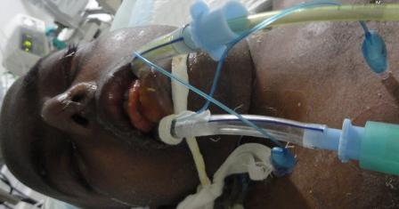
A 22 years old male, suffering from hepatic encephalopathy without coagulopathy, presented in our emergency department (ED) with Glasgow Coma Scale (GCS) 7/15 at midnight. Lung auscultation revealed right sided crepitations with SpO2 of 82% (probably due to aspiration). He required intubation for airway protection to prevent further aspiration and maintenance of oxygenation. His airway examination was apparently normal with unknown fasting status. After pre-oxygenation for 3 minutes he was induced with fentanyl and midazolam and preservative free lignocaine. On laryngoscopy, just tip of the epiglottis was barely visible (CL grade 3 b), even after optimum external laryngeal manipulation. An 8 mm endotracheal tube (ETT) with stylet was tried and the cuff inflated with air, but five point auscultation confirmed it to be inside the esophagus. We passed a 14 F nasogastric tube through the esophageal ETT in situ and suctioned his stomach contents. Then shifting the first ETT to left corner of the patient’s mouth, a second laryngoscopy was done. A sterile 9 F AC (Medtronic, Minnesota, USA) with guide wire, lubricated with lignocaine jelly, was passed just below the tip of epiglottis and through the glottis; a tactile sensation was felt by keeping the palm on trachea. A second ETT was now railroaded over the AC (Figure 1). In this way, the first ETT in the esophagus helped in preventing aspiration and guiding AC into trachea. Whole of the procedure was completed within 4 minutes without desaturating the patient (Figure 2).
Incidence of difficult laryngoscopy (defined as tracheal intubation requiring >2 attempts at direct laryngoscopy) is 1.5-8.5% in general anesthesia with failed intubation 0.3%.1 Alternative airway devices like the rigid or flexible fibreoptic bronchoscope, intubating laryngeal mask airway (ILMA), videolaryngoscope, C-Trach, and light wand etc. are very effective in this situation. Airway management in ED is often more challenging than in the operating rooms due to lack of difficult airway gadgets and expert help. In unanticipated difficult airway in pre-hospital emergency setting, people have used gum elastic bougies, ILMA and cricothyroidotomy.2] In the absence of such airway instruments in ED, an our simple technique may be tried. We suggest that if an esophageal intubation has inadvertently been done in a difficult intubation situation, don’t remove it, rather keep it inflated to prevent aspiration. Even mask ventilation can be successfully done with the help of a small gauge piece sealing on left angle of the mouth. We used a sterilised AC with atraumatic guide wire with minimum cost. AC has been used for nasogastric tube insertion but never used in airway management.3,4 AC can also be resterilised and can be kept inside the resuscitation kit from cardiac catheterization laboratories.
REFERENCES PubMed
1. Crosby ET, Cooper RM, Douglas MJ, Doyle DJ, Hung OR, Labrecque P, et al. The unanticipated difficult airway with recommendations for management. Can J Anaesth. 1998;45:757-76. [PubMed]
2. Combes X, Jabre P, Margenet A, Merle JC, Leroux B, Dru M, et al. Unanticipated Difficult Airway Management in the Prehospital Emergency Setting: Prospective Validation of an Algorithm. Anesthesiology. 2011;114:105-10. [PubMed] [Free Full Text]
3. Rutala WA, Weber DJ. Healthcare infection control practices advisory committee (HICPAC). Guideline for disinfection and sterilization in healthcare facilities, 2008. Available from: http://www.cdc.gov/hipac/pdf/guidelines/Disinfection_Nov_2008.pdf. [Online Access].
4. Ghatak T, Samanta S, Baronia AK . A new technique to insert nasogastric tube in an unconscious intubated patient. North Am J Med Sci. 2013;5:68-70. [PubMed] [Free Full Text]
Figure 1: Angiographic catheter with guide wire as airway exchange through ETT
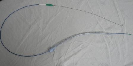
Figure 2: Patient with both ETT (esophageal & tracheal) in situ.