
Divanshu Gupta, Alaka Purohit
Department of Anesthesiology, Sawai Man Singh (SMS) Medical College, Jaipur, Rajasthan, (India)
Correspondence: Dr. Divanshu Gupta, Department of Anesthesiology, Sawai Man Singh (SMS) Medical College, Jawaharlal Nehru Marg, Gangawal Park, Jaipur, Rajasthan 302004, (India); E-mail: divanshu2387@yahoo.com; Phone: 9887177365
ABSTRACT
Osteogenesis imperfecta (OI) is a rare, genetically inherited syndrome involving connective tissue, resulting in anatomic and physiologic abnormalities, which may present with challenges during anesthesia. We hereby report a case of OI type I, who presented with distinctively blue sclerae, hearing loss, kyphoscoliosis and mild pulmonary restrictive disease. She was scheduled to have Rush nail removal from her femurs.
Key words: Osteogenesis imperfecta; Anesthesia, Spinal
Citation: Gupta D, Purohit A. Spinal anesthesia in osteogenesis imperfecta. Anaesth Pain & Intensive Care 2016;20(1):86-88
INTRODUCTION
Osteogenesis imperfecta (OI) also known as Brittle bone disease, Lobstein’s disease, fragilis Ossium, osteopsathyrosis idiopathica or Vrolik’s disease. Vrolik was the first to use the term osteogenesis imperfecta. OI is an inherited disease of connective tissue that affects bone, sclerae and the inner ear, caused by a mutation of the collagen type I – COLIA 1 and COLIA 2 genes.1
According to SILLENCE Classification disease has been classified into four types;2
TYPE I (the commonest form) is inherited as an autosomal dominant fashion and is characterized by blue sclerae, fragile bones, hyperextensible joints, progressive hearing loss and dentinogenesis imperfecta. Type II is lethal either in utero or in the perinatal period. Type I and Type IV are inherited as an autosomal dominant fashion, while Type II and III are autosomal recessive. In Type III, patients usually die in childhood or adolescence as a consequence of cardiopulmonary complications. In Type IV patient has skeletal deformities without ocular, audiological, and dental abnormalities.2
The defect in skeletal growth is a result of lack of normal ossification of endochondrial bone resulting in increased fragility of bones, hypermobile limbs, and kyphoscoliosis. In such disorder anesthesia challenges are a difficult airway, short neck, risk of odontoaxial dislocation, and cervical vertebrae, mandible and tooth fractures during laryngoscopy and intubation. These patients may also have cardiac valvular lesions, cor pulmonale, neurologic abnormalities, hyperhydrosis, cleft palate, metabolic abnormalities, malignant hyperthermia, obstructive uropathy following renal and ureteric stones and platelet dysfunction.1
We report a case of anesthetic management of a known case of OI Type I, who underwent a rush nail removal of femur.
CASE REPORT
A 15 year old female presented in pre anesthetic clinic for rush nail removal of femur. She was a known case of OI Type I, and had a history of recurrent fractures for which she had been operated thrice before uneventfully under general anesthesia. There was no history of OI in the family.
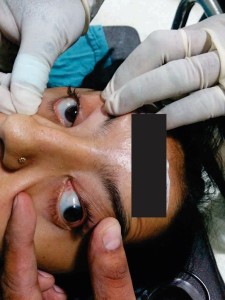
Figure 1: Blue colored sclera in OI
On general examination, she was short statured with a height of 103 cm, weighing 49 kg, blue sclerae, fragile bones, kyphoscoliosis and progressive hearing loss. Her blood pressure was 130/80 mmHg, heart rate was 80 beats/min. There was no pallor, icterus, cyanosis, clubbing, lymphadenopathy, or pedal edema. Respiratory system revealed barrel shaped chest with equal bilateral air entry. Cardiovascular system revealed normal heart sounds. ECG, liver and kidney function tests, and the coagulation profile were normal. Chest x-ray revealed mediastinal widening. X-ray spine revealed dorsolumber kyphoscoliosis to left. Pulmonary function tests suggested mild restrictive disease. Airway assessment showed acceptable flexion and extension at neck with interincisor gap 3 cm and normal dentition; she was mallampati class III. The patient was accepted for surgery as ASA grade III. In view of risk of odontoaxial dislocation and a normal coagulation profile, we decided to proceed with a spinal anesthetic technique. Thorough operating room preparations were completed including difficult airway equipment and measures to deal with hyperthermia if any.
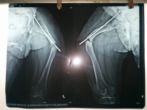
Figure 2: Showing osteolysis and fractures
The patient was positioned very carefully on the operating table, pressure points were padded adequately. An 18G IV cannula was inserted on the dorsum of the right hand. Routine monitors were attached to the patient.
Her baseline blood pressure was 128/80 mmHg, heart rate was 90 beats/min, SpO2 on room air was 98%. A preload of 500 ml Ringer’s lactate was infused. Despite the kyphoscoliosis no difficulty was experienced in finding the subarachnoid space, and spinal injection of bupivacaine heavy 0.5 % 2.5 ml was given in the sitting position in L3 – L4 space with a 25G quincke needle. The motor block was upto T 10 level. The surgery lasted for one hour and 45 min. There was no significant variation in the vital signs, and the recovery was uneventful.
DISCUSSION
OI is a rare autosomal dominant inherited disease of connective tissue that affects bones, sclerae, and inner ears. It affects 6-7 of 100,000 people and occurs in 1: 20000 births.3
Anesthetic management of OI is influenced by diversity of presentation viz. coexisting orthopedic deformities, fragile bones prone for fractures at the time of positioning, platelet dysfunction, cardiovascular abnormalities like mitral valve prolapse, tendency to develop malignant hyperthermia, anticipated difficult intubation because of abnormal cervical spine mobility, fragile teeth, odontoaxial dislocation, and risk of mandibular and facial fractures.4
There have been several case reports of managing such cases under general anesthesia. Karabiyk et al recommended TIVA along with intubating LMA,4 while Malde et al used balanced anesthesia in a case of OI with gross deformity of the pelvis for abdominal hysterectomy,5 though Sachin et al observed a significant degree of movement between first and second cervical vertebra during direct laryngoscopy and with use of the intubating LMA.6
Best technique in these patients is probably spinal anesthesia as it avoids the need of tracheal intubation, reduces the risk of malignant hyperthermia, and facilitates early detection of thyroid storm. Hence, we preferred spinal anesthesia, as patient was undergoing lower limb surgery, and we wanted to avoid ventilation perfusion mismatch likely to occur in general anesthesia due to spine and chest deformities affecting vital capacity and chest compliance. Moreover, succinyl choline induced fasciculations could lead to fractures, inhalational agents also poses risk for malignant hyperthermia. Though general consensus is that hyperthermia in such cases is due to hypermetabolic state and is not of the malignant type.
Automated blood pressure cuffs are hazardous as overinflation might result in fractures, so either invasive blood pressure or manual sphygmomanometer should be used. Pressure points were well padded. Thorough preoperative workup including patient coagulation profile, cardiorespiratory workup including electrocardiography, echocardiography, pulmonary function tests are mandatory.
CONCLUSION
Patients with OI pose a significant challenge to anesthesiologist owing to difficult airway and associated multiple comorbidities, a meticulous workup of the patient is required. Regional anesthetic techniques should be preferred wherever permissible to have favorable outcome.
Regional techniques appear to be safe in the hands of an experienced and vigilant anesthesiologist.
Conflict of interest: None
Author contribution: DG: Follow up of the case, manuscript writing; AP: Manuscript editing
REFERENCES
- Bhandari G, Shahi KS, Bhadoria P, Bhalotra AR, Sandhya OD, Arya M. Osteogenesis imperfecta : no place for imperfect anaesthesiologist. Indian J Anaesth 2008;52(5):577-580. [Free full text]
- Gornik-Właszczuk E, Majewski J, Szczygieł R, Dusiel A. Anaesthesia in children with osteogenesis imperfecta— report of 14 general anaesthetics in three children. Anaesthesiol Intensive Ther. 2013 Apr-Jun;45(2):85–88. [PubMed] [Free full text]
- Bhardwaj M, Kaur K, Johar S, Ruchi S, Hooda S.Anaesthetic management in a patient with osteogenesis imperfecta and a fractured femur. South Afr J Anaesth Analg 2014;20(2):132-135.
- Karabiyik L, Parpucu M, Kurtipek O. Total intravenous anaesthesia and the use of an intubating laryngeal mask in a patient with osteogenesis imperfecta. Acta Anaesthesiol Scand. 2002 May;46(5):618-19. [PubMed]
- Malade AD, Jagtap SR, Pantvaidya SH, Kenkare JS. Osteogenesis Imperfecta: Anaesthetic management of a patient for abdominal hysterectomy (a case report). Indian J Anaesth 1993;41:203-06.
- Sachin A, Salman MA, Erden IA, Aypar U. Upper cervical vertebral movement during intubating laryngeal mask, fibreoptic and direct laryngeoscopy: A video fluoroscopic study. Eur J Anaesthesiol 2004;21:819-23.
