Bahtiar Ahmad 1 , Kohar Hari Santoso 2 , Arie Utarani 3 , Elizeus Hanindito 4 , Bambang Pujo Semedi 5
Author afiliations:
Background: Congenital cervical teratoma is a rare tumor in neonates that can cause life-threatening airway obstruction. Early diagnosis and airway planning are essential to improve outcomes. Anesthetic management poses significant challenges due to airway deviation and limited visualization during intubation.
Case: We report a case of a 7-month-old male infant with a large cervical teratoma (10.5 × 10.8 × 10.7 cm). Anesthesia was induced using sevoflurane with spontaneous ventilation. Direct laryngoscopy was performed using a Magill laryngoscope while an assistant repositioned the mass to expose the trachea. Intubation was successful on the first attempt using a 4.5 mm ETT. Backup airway tools, including a fiberoptic laryngoscope and LMA, were prepared but not required. Surgery lasted 360 minutes, with minimal blood loss and stable intraoperative hemodynamics. The patient was transferred to the PICU postoperatively.
Discussion: This case highlights the importance of early planning, maintaining spontaneous ventilation, and preparing for difficult airway scenarios in pediatric cervical teratoma. Compared to previous reports, the success of first-attempt intubation and minimal complications highlight the value of manual mass manipulation and adequate preparation.
Conclusion: Airway management in congenital cervical teratoma requires careful preoperative evaluation, availability of advanced airway tools, and multidisciplinary coordination. Successful outcomes depend on preserving airway patency, ensuring optimal ventilation, and anticipating complications with thorough preparation.
Keywords: anesthesia; intubation; ventilation; management; congenital cervical teratoma;pediatric
Citation: Ahmad B, Santoso KH, Utarani A, Hanindito E. Semedi BP. Anesthetic management of a patient with difficult intubation and ventilation due to a congenital cervical teratoma. Anaesth. pain intensive care 2026;30(6):780-783. DOI: 10.35975/apic.v30i6.3275
Received: September 14, 2025; Revised: October 26, 2025; Accepted: June 02, 2026
Cervical teratomas are rare congenital tumors that originate from all three germinal layers and have the potential to cause airway obstruction. Despite their low incidence of approximately 1 in 20,000-40,000 live births.12 These lesions typically present as large cervical masses that can exert significant compression on the upper airway, leading to respiratory distress or even life-threatening obstruction at birth or during infancy. Despite histologically benign features in most cases, the clinical management of cervical teratomas remains challenging due to their size, vascularity, and anatomic location near vital structures such as the trachea and major cervical vessels.2–4
Prenatal detection via ultrasonography or magnetic resonance imaging occurs in only about 20% of cases, limiting the opportunity for planned interventions such as EXIT (Ex-Utero Intrapartum Treatment) procedures to secure the airway before cord clamping. In unexpected postnatal presentations, immediate airway stabilization becomes paramount, often requiring a combination of techniques including jaw thrust, bag-mask ventilation, direct laryngoscopy, or emergent tracheostomy. Anesthetic and airway management in these patients is particularly challenging. Pediatric airway anatomy itself poses inherent difficulties: a proportionally larger tongue, a more cephalad and anterior larynx (C2–C3), a narrower subglottic diameter, and increased airway compliance.5When compounded by a massive cervical teratoma, these features create an extremely difficult airway scenario. Various approaches have been described, including inhalational induction with preserved spontaneous breathing, fiberoptic intubation, videolaryngoscopy, or emergency tracheostomy.6
This case highlights the perioperative management of a 7-month-old infant with a large congenital cervical teratoma, focusing on strategies for safe intubation, ventilation, and perioperative monitoring. By comparing with previous reports, this case underscores both the common challenges and unique adaptations made in resource-limited but real-world clinical contexts.
A 7-month-old male infant with a congenital cervical teratoma measured 10.5 × 10.8 × 10.7 cm. The patient's general condition is good with stable vital signs. Anesthesia problems in this patient are pediatric, as is the potential for difficult intubation and ventilation. Anaesthesia was induced with sevoflurane in oxygen until an adequate level of anesthesia with spontaneous ventilation was achieved. Direct laryngoscopy was performed with Magill’s laryngoscope while an assistant pulled a mass to the posterolateral to free up the trachea. A fiberoptic laryngoscope, LMA, and boogie were prepared if the intubation attempt failed. The higher laryngeal position (C2-C3) makes visualization during laryngoscopy more difficult, and a larger tongue relative to the oral cavity further complicates access to the vocal cords. The trachea was intubated with a size 4.5 mm ETT in the first attempt. EtCO2 was maintained within 28–35 mmHg, and an arterial line was inserted to monitor invasive blood pressure. A 3-way infusion was also performed for drug access and blood transfusion. A procedure runs for 360 minutes. The duration of anesthesia is 420 minutes. Blood loss of 50 mL. Urine output of 15 mL. Postoperatively, the patient was taken to the PICU for intensive care.
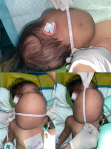
Figure 1: Neck mass measurement before surgery.
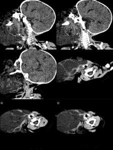
Figure 2: Head and Neck CT Scan.
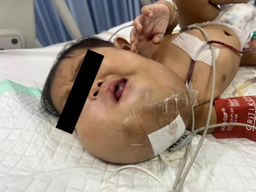
Figure 3: After surgery.
This case highlights the perioperative management of a 7-month-old infant with a large congenital cervical teratoma, focusing on strategies for safe intubation, ventilation, and perioperative monitoring. By comparing with previous reports, this case underscores both the common challenges and unique adaptations made in resource-limited but real-world clinical contexts. Airway management in patients with congenital cervical teratomas is a major challenge for anesthesiologists, especially in pediatric patients.5 This case highlights the difficulties in managing the airway in a pediatric patient with a large cervical teratoma, with emphasis on the first successful intubation using the Magill laryngoscope after manual repositioning of the tumor. Compared to the existing literature, this case involved a 7-month-old infant boy with a teratoma measuring 10.5 × 10.8 × 10.7 cm, while the report of Jain & Varshney (2013) described a 3-year-old girl with a larger teratoma (18 × 12 cm) that had been growing for two years, causing severe tracheal deviation and intubation difficulties. 7. Inhalation induction with sevoflurane while preserving spontaneous breathing is recommended to avoid complete airway collapse that may occur with neuromuscular blockade. 5
Residual mass approximately 10.5 × 10.8 × 10.7 cm (previously: 6.8 × 7.1 × 9.8 cm) in the right cervical region with the above-described extension, representing congenital cervical teratoma with feeding vessels from he right lingual artery, right facial artery, and right ascending pharyngeal artery. The mass has enlarged compared to previous examination. No intralesional hemorrhage seen.
Recent literature provides additional insights into strategies for managing such complex airway cases. Fiberoptic intubation has been reported as a gold standard in difficult neonatal airways, often facilitated through a laryngeal mask airway (LMA) used as a conduit. 9,10 The LMA itself is widely recognized as a valuable rescue device in cases of failed intubation, allowing temporary ventilation and providing a channel for fiberoptic intubation.
Total intraoperative urine output was 15 mL, with an estimated blood loss of 50 mL. The patient was extubated fully conscious at 17:00.
Videolaryngoscopy has also gained increasing importance, with devices such as Airtraq and C-MAC demonstrating improved visualization of the glottis in cases where direct laryngoscopy is obstructed by cervical masses.11 These modern technologies complement traditional techniques, improving first-attempt success rates and patient safety.
Intraoperative management should also address the potential for significant blood loss given the vascularity of teratomatous tissue. Our case involved minimal hemorrhage (50 mL) due to careful dissection and vigilant hemodynamic monitoring with invasive arterial blood pressure and end-tidal CO₂ (EtCO₂) surveillance. Other series have documented the role of preoperative embolization to reduce intraoperative transfusion requirements in exceptionally vascular tumors, although this was not necessary in our instance.2,7
This discrepancy emphasizes the importance of early diagnosis, meticulous airway planning and availability of advanced airway devices, especially in cases with severe tracheal deviation. Postoperative care in the pediatric intensive care unit (PICU) is crucial for continued airway observation and support. Late airway complications such as tracheomalacia or recurrent laryngeal nerve palsy have been reported, necessitating ongoing multidisciplinary follow-up. 5
Taken together, this case reinforces the importance of multidisciplinary collaboration, early planning, and adaptability in resource-limited environments. It also contributes to the literature by demonstrating that manual mass manipulation during laryngoscopy, although rarely described, can be a valuable adjunct to optimize glottic exposure in selected patients.
Anesthetic management of cervical teratoma requires thorough airway evaluation preoperatively as well as imaging to anticipate potential complications. Careful preparation for airway challenges is essential, including the availability of advanced equipment and adequate surgical support. In our case, an effective intubation strategy with a Magill laryngoscope after manual repositioning of the tumor proved successful in overcoming tracheal obstruction. This highlights the fact that even in resource-constrained environments, careful preparation and adaptive techniques can achieve safe outcomes. Recent reports confirm that timely airway stabilization, followed by early tumor resection, leads to favorable survival and developmental outcomes.12,13
Ultimately, successful management of congenital cervical teratomas is rooted in early recognition, multidisciplinary collaboration, and flexible airway strategies informed by both modern technology and practical bedside adaptations. By integrating evidence from global literature and local experience, this case underscores how comprehensive planning and innovative techniques remain the cornerstones of improving perioperative safety and long-term prognosis in children with this rare but high-risk condition.
5. Conflict of interest
The authors declare no conflict of interest.
6. Funding
The study utilized the hospital resources only, and no external or industry funding was involved.
7. Author contribution
Both authors took equal part of the case report.
Author afiliations:
- Bahtiar Ahmad, Department of Anesthesiology, Faculty of Medicine, Airlangga University, Surabaya, Indonesia
- Kohar Hari Santoso, Department of Anesthesiology, Faculty of Medicine, Airlangga University, Surabaya, Indonesia
- Arie Utarani, Department of Anesthesiology, Faculty of Medicine, Airlangga University, Surabaya, Indonesia
- Elizeus Hanindito, Department of Anesthesiology, Faculty of Medicine, Airlangga University, Surabaya, Indonesia
- Bambang Pujo Semedi, Department of Anesthesiology, Faculty of Medicine, Airlangga University, Surabaya, Indonesia
ABSTRACT
Background: Congenital cervical teratoma is a rare tumor in neonates that can cause life-threatening airway obstruction. Early diagnosis and airway planning are essential to improve outcomes. Anesthetic management poses significant challenges due to airway deviation and limited visualization during intubation.
Case: We report a case of a 7-month-old male infant with a large cervical teratoma (10.5 × 10.8 × 10.7 cm). Anesthesia was induced using sevoflurane with spontaneous ventilation. Direct laryngoscopy was performed using a Magill laryngoscope while an assistant repositioned the mass to expose the trachea. Intubation was successful on the first attempt using a 4.5 mm ETT. Backup airway tools, including a fiberoptic laryngoscope and LMA, were prepared but not required. Surgery lasted 360 minutes, with minimal blood loss and stable intraoperative hemodynamics. The patient was transferred to the PICU postoperatively.
Discussion: This case highlights the importance of early planning, maintaining spontaneous ventilation, and preparing for difficult airway scenarios in pediatric cervical teratoma. Compared to previous reports, the success of first-attempt intubation and minimal complications highlight the value of manual mass manipulation and adequate preparation.
Conclusion: Airway management in congenital cervical teratoma requires careful preoperative evaluation, availability of advanced airway tools, and multidisciplinary coordination. Successful outcomes depend on preserving airway patency, ensuring optimal ventilation, and anticipating complications with thorough preparation.
Keywords: anesthesia; intubation; ventilation; management; congenital cervical teratoma;pediatric
Citation: Ahmad B, Santoso KH, Utarani A, Hanindito E. Semedi BP. Anesthetic management of a patient with difficult intubation and ventilation due to a congenital cervical teratoma. Anaesth. pain intensive care 2026;30(6):780-783. DOI: 10.35975/apic.v30i6.3275
Received: September 14, 2025; Revised: October 26, 2025; Accepted: June 02, 2026
1. INTRODUCTION
Cervical teratomas are rare congenital tumors that originate from all three germinal layers and have the potential to cause airway obstruction. Despite their low incidence of approximately 1 in 20,000-40,000 live births.12 These lesions typically present as large cervical masses that can exert significant compression on the upper airway, leading to respiratory distress or even life-threatening obstruction at birth or during infancy. Despite histologically benign features in most cases, the clinical management of cervical teratomas remains challenging due to their size, vascularity, and anatomic location near vital structures such as the trachea and major cervical vessels.2–4
Prenatal detection via ultrasonography or magnetic resonance imaging occurs in only about 20% of cases, limiting the opportunity for planned interventions such as EXIT (Ex-Utero Intrapartum Treatment) procedures to secure the airway before cord clamping. In unexpected postnatal presentations, immediate airway stabilization becomes paramount, often requiring a combination of techniques including jaw thrust, bag-mask ventilation, direct laryngoscopy, or emergent tracheostomy. Anesthetic and airway management in these patients is particularly challenging. Pediatric airway anatomy itself poses inherent difficulties: a proportionally larger tongue, a more cephalad and anterior larynx (C2–C3), a narrower subglottic diameter, and increased airway compliance.5When compounded by a massive cervical teratoma, these features create an extremely difficult airway scenario. Various approaches have been described, including inhalational induction with preserved spontaneous breathing, fiberoptic intubation, videolaryngoscopy, or emergency tracheostomy.6
This case highlights the perioperative management of a 7-month-old infant with a large congenital cervical teratoma, focusing on strategies for safe intubation, ventilation, and perioperative monitoring. By comparing with previous reports, this case underscores both the common challenges and unique adaptations made in resource-limited but real-world clinical contexts.
2. CASE REPORT
A 7-month-old male infant with a congenital cervical teratoma measured 10.5 × 10.8 × 10.7 cm. The patient's general condition is good with stable vital signs. Anesthesia problems in this patient are pediatric, as is the potential for difficult intubation and ventilation. Anaesthesia was induced with sevoflurane in oxygen until an adequate level of anesthesia with spontaneous ventilation was achieved. Direct laryngoscopy was performed with Magill’s laryngoscope while an assistant pulled a mass to the posterolateral to free up the trachea. A fiberoptic laryngoscope, LMA, and boogie were prepared if the intubation attempt failed. The higher laryngeal position (C2-C3) makes visualization during laryngoscopy more difficult, and a larger tongue relative to the oral cavity further complicates access to the vocal cords. The trachea was intubated with a size 4.5 mm ETT in the first attempt. EtCO2 was maintained within 28–35 mmHg, and an arterial line was inserted to monitor invasive blood pressure. A 3-way infusion was also performed for drug access and blood transfusion. A procedure runs for 360 minutes. The duration of anesthesia is 420 minutes. Blood loss of 50 mL. Urine output of 15 mL. Postoperatively, the patient was taken to the PICU for intensive care.
Figure 1: Neck mass measurement before surgery.
Figure 2: Head and Neck CT Scan.
Figure 3: After surgery.
3. DISCUSSION
This case highlights the perioperative management of a 7-month-old infant with a large congenital cervical teratoma, focusing on strategies for safe intubation, ventilation, and perioperative monitoring. By comparing with previous reports, this case underscores both the common challenges and unique adaptations made in resource-limited but real-world clinical contexts. Airway management in patients with congenital cervical teratomas is a major challenge for anesthesiologists, especially in pediatric patients.5 This case highlights the difficulties in managing the airway in a pediatric patient with a large cervical teratoma, with emphasis on the first successful intubation using the Magill laryngoscope after manual repositioning of the tumor. Compared to the existing literature, this case involved a 7-month-old infant boy with a teratoma measuring 10.5 × 10.8 × 10.7 cm, while the report of Jain & Varshney (2013) described a 3-year-old girl with a larger teratoma (18 × 12 cm) that had been growing for two years, causing severe tracheal deviation and intubation difficulties. 7. Inhalation induction with sevoflurane while preserving spontaneous breathing is recommended to avoid complete airway collapse that may occur with neuromuscular blockade. 5
Residual mass approximately 10.5 × 10.8 × 10.7 cm (previously: 6.8 × 7.1 × 9.8 cm) in the right cervical region with the above-described extension, representing congenital cervical teratoma with feeding vessels from he right lingual artery, right facial artery, and right ascending pharyngeal artery. The mass has enlarged compared to previous examination. No intralesional hemorrhage seen.
Recent literature provides additional insights into strategies for managing such complex airway cases. Fiberoptic intubation has been reported as a gold standard in difficult neonatal airways, often facilitated through a laryngeal mask airway (LMA) used as a conduit. 9,10 The LMA itself is widely recognized as a valuable rescue device in cases of failed intubation, allowing temporary ventilation and providing a channel for fiberoptic intubation.
Total intraoperative urine output was 15 mL, with an estimated blood loss of 50 mL. The patient was extubated fully conscious at 17:00.
Videolaryngoscopy has also gained increasing importance, with devices such as Airtraq and C-MAC demonstrating improved visualization of the glottis in cases where direct laryngoscopy is obstructed by cervical masses.11 These modern technologies complement traditional techniques, improving first-attempt success rates and patient safety.
Intraoperative management should also address the potential for significant blood loss given the vascularity of teratomatous tissue. Our case involved minimal hemorrhage (50 mL) due to careful dissection and vigilant hemodynamic monitoring with invasive arterial blood pressure and end-tidal CO₂ (EtCO₂) surveillance. Other series have documented the role of preoperative embolization to reduce intraoperative transfusion requirements in exceptionally vascular tumors, although this was not necessary in our instance.2,7
This discrepancy emphasizes the importance of early diagnosis, meticulous airway planning and availability of advanced airway devices, especially in cases with severe tracheal deviation. Postoperative care in the pediatric intensive care unit (PICU) is crucial for continued airway observation and support. Late airway complications such as tracheomalacia or recurrent laryngeal nerve palsy have been reported, necessitating ongoing multidisciplinary follow-up. 5
Taken together, this case reinforces the importance of multidisciplinary collaboration, early planning, and adaptability in resource-limited environments. It also contributes to the literature by demonstrating that manual mass manipulation during laryngoscopy, although rarely described, can be a valuable adjunct to optimize glottic exposure in selected patients.
4. CONCLUSION
Anesthetic management of cervical teratoma requires thorough airway evaluation preoperatively as well as imaging to anticipate potential complications. Careful preparation for airway challenges is essential, including the availability of advanced equipment and adequate surgical support. In our case, an effective intubation strategy with a Magill laryngoscope after manual repositioning of the tumor proved successful in overcoming tracheal obstruction. This highlights the fact that even in resource-constrained environments, careful preparation and adaptive techniques can achieve safe outcomes. Recent reports confirm that timely airway stabilization, followed by early tumor resection, leads to favorable survival and developmental outcomes.12,13
Ultimately, successful management of congenital cervical teratomas is rooted in early recognition, multidisciplinary collaboration, and flexible airway strategies informed by both modern technology and practical bedside adaptations. By integrating evidence from global literature and local experience, this case underscores how comprehensive planning and innovative techniques remain the cornerstones of improving perioperative safety and long-term prognosis in children with this rare but high-risk condition.
5. Conflict of interest
The authors declare no conflict of interest.
6. Funding
The study utilized the hospital resources only, and no external or industry funding was involved.
7. Author contribution
Both authors took equal part of the case report.
8. REFERENCES
- Jordan RB, Gauderer MWL. Cervical teratomas: An analysis. Literature review and proposed classification. J Pediatr Surg. 1988 Jun;23(6):583–91.
- Hochwald O, Gil Z, Gordin A, Winer Z, Avrahami R, Abargel E, et al. Three-step management of a newborn with a giant, highly vascularized, cervical teratoma: A case report. J Med Case Rep. 2019 Mar 10;13(1).
- Siverio N H, Fernández LT, González A, Jm TL. Hernández Siverio N et al Giant Congenital Cervical Teratoma: Diagnosis, Management and Long-term Follow-up.
- Mohanty M, Sahu P, Jaiswal A, Singal R, Gupta S, Kohli G, et al. A huge immature cervical teratoma; Antenatal diagnosis, and its management - An unusual entity. J Clin Neonatol. 2013;2(1):42.
- Neidich MJ, Prager JD, Clark SL, Elluru RG. Comprehensive Airway Management of Neonatal Head and Neck Teratomas. Otolaryngology–Head and Neck Surgery. 2011 Feb 4;144(2):257–61.
- Jain G, Varshney R. Anaesthetic challenges in a patient presenting with huge neck teratoma. Saudi J Anaesth. 2013;7(2):210.
- Jain G, Varshney R. Anaesthetic challenges in a patient presenting with huge neck teratoma. Saudi J Anaesth. 2013 Apr;7(2):210–2.
- Kwari YD, Sani A, Balami AP. Management of difficult airways due to cervical teratoma presenting with respiratory distress at birth: Reports of two cases. J Anesth Clin Res. 2012 Feb;3(2).
- Ramesh A, Gupta KK, Deep G, Singh A. Airway management of postburn neck contracture in pediatric patient – A challenge for anesthesiologist! Saudi J Anaesth. 2024 Apr;18(2):286–9.
- Jain G, Varshney R. Anaesthetic challenges in a patient presenting with huge neck teratoma. Saudi J Anaesth. 2013;7(2):210.
- Prakash S, Rapsang AG, Mahajan S, Bhattacharjee S, Singh R, Gogia AR. Comparative Evaluation of the Sniffing Position with Simple Head Extension for Laryngoscopic View and Intubation Difficulty in Adults Undergoing Elective Surgery. Anesthesiol Res Pract. 2011;2011:1–6.
- Elmasalme F, Giacomantonio M, Clarke K, Othman E, Matbouli S. Congenital Cervical Teratoma in Neonates Case Report and Review. European Journal of Pediatric Surgery. 2000 Aug 25;10(04):252–7.
- Ali IM, Yaşar M, Abdi AA, Dırken ES. Successful surgıcal management of a giant neck teratoma in a newborn Baby: A case report. Annals of Medicine & Surgery. 2022 Oct;82.