Theresia Chandra Tania Novy1, Christopher Lampah2, Dewa Gede Satria Ambara Putra3, Daniel Hadimartana4, Maria Jessica Yaputri5
Authors affiliation:
Introduction: Osteoarthritis (OA) is a progressive degenerative joint disease commonly affecting weight-bearing joints such as the knee and hip, leading to pain, stiffness, and functional impairment. Conventional treatments often provide only temporary symptom relief, prompting increasing interest in regenerative approaches such as mesenchymal stem cell therapy.
Case Report: We report a 60-year-old woman with a one-year history of left hip and knee pain refractory to conservative management. Clinical and imaging findings were consistent with osteoarthritis and suspected avascular necrosis of the left femoral head. Initial ultrasound examination demonstrated intra-articular effusion in both joints. The patient first underwent ultrasound-guided intra-articular corticosteroid injection followed by viscosupplementation. Due to persistent symptoms, regenerative therapy using umbilical cord–derived mesenchymal stem cells (UC-MSCs) was performed under ultrasound guidance. A total of 70 × 10⁶ cells were injected intra-articularly, including 20 × 10⁶ cells into each knee and 30 × 10⁶ cells into the left hip. Progressive improvement in pain and function was observed without significant adverse events.
Conclusion: Ultrasound-guided intra-articular UC-MSC therapy may represent a promising minimally invasive treatment option for patients with osteoarthritis who do not respond adequately to conventional therapy.
Keywords: hip osteoarthritis; knee osteoarthritis; mesenchymal stem cells; regenerative therapy; ultrasound-guided injection
Citation: Novy TCT, Lampah C, Putra DGS, Hadimartana D, Yaputri MJ. Ultrasound-guided stem cell therapy for hip and knee osteoarthritis: a case report. Anaesth. pain intensive care 2026;30(6):773-779.
DOI: 10.35975/apic.v30i6.3274
Received: xxxxxxxxx; Revised: xxxxxxxxxxx; Accepted: xxxxxxxxxx
Osteoarthritis (OA) is the most prevalent degenerative joint disorder, characterized by chondrocyte damage caused by inflammatory cytokines and mechanical stress, leading to joint space narrowing, osteophyte formation, and subchondral bone changes. Globally, approximately 240 million adults suffer from symptomatic OA, and its prevalence continues to increase with aging populations. The main symptoms include pain, stiffness, and limited joint motion. Risk factors include genetics, sex, mechanical stress, and previous trauma, with the knee most commonly affected, followed by the hip.1 Diagnosis is usually clinical, although radiographic grading systems such as Tönnis and Kellgren–Lawrence are widely used to assess disease severity.2 Current management includes weight reduction, physiotherapy, pharmacologic therapy, and intra-articular injections, while joint arthroplasty remains invasive and associated with potential complications.3 Mesenchymal stem cells (MSCs) have emerged as a promising regenerative therapy due to their immunomodulatory, anti-inflammatory, and cartilage-regenerative potential.⁴ This case report aims to present a regenerative intervention using umbilical-derived stem cells in a 60-year-old woman with hip and knee OA.
We report a 60-year-old woman presenting with a one-year history of left buttock and hip pain with a Numeric Rating Scale (NRS) score of 8. The pain was aggravated by standing up and sitting down and was accompanied by mild left knee pain during weight-bearing activities such as standing and walking. The patient had previously consulted an orthopaedic specialist and a neurologist and received pharmacological therapy, but without significant improvement.
The pain later recurred, predominantly involving the left knee and thigh, particularly when rising from bed or changing position from sitting to standing. She also reported sleep disturbances caused by pain during positional changes. Her daily activities included running a shop and baking, which required prolonged standing, as well as exercising on a stationary bicycle.
Physical examination revealed an antalgic gait and tenderness over the right hip joint. Provocative low back maneuvers on the left side were positive, suggesting hip osteoarthritis, left piriformis syndrome, and left sacroiliitis. Examination of the left knee demonstrated joint crepitus and mild tenderness over the suprapatellar bursa, medial collateral ligament (MCL), and pes anserine bursa, suggesting bursitis and knee osteoarthritis as possible differential diagnoses.
Radiographic evaluation of both knees and the left hip joint is shown in Figure 1. Knee radiographs demonstrated osteophyte formation consistent with mild bilateral knee osteoarthritis (Figure 1A–B), while hip radiography revealed degenerative changes in the left hip joint (Figure 1C). Previous MRI findings further indicated suspected avascular necrosis (AVN) of the left femoral head with secondary osteoarthritis of the left hip and a superior labral tear. A cystic lesion containing both fat and solid components was also observed in the left pelvic cavity. MRI images of the pelvis and hip region are presented in Figure 1D–E.
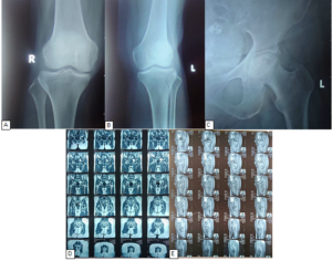
Figure 1. Radiographic and MRI findings of the knee and hip joints. (A) Anteroposterior radiograph of the right knee demonstrating osteophyte formation consistent with mild osteoarthritis. (B) Anteroposterior radiograph of the left knee showing similar osteoarthritic changes. (C) Anteroposterior radiograph of the left hip revealing degenerative changes suggestive of secondary osteoarthritis. (D–E) Magnetic resonance imaging (MRI) of the pelvis and hip region demonstrating suspected avascular necrosis (AVN) of the left femoral head with associated secondary osteoarthritis and a superior labral tear. A cystic lesion containing both fat and solid components is also observed within the left pelvic cavity.
At the initial visit, ultrasound examination demonstrated joint effusion and osteophytes in the hip and knee joints. Quantitative measurements of intra-articular effusion are summarized in Table 1, which showed greater effusion in the left hip joint and mild effusion in both knees. Ultrasound-guided intra-articular injection with triamcinolone was subsequently performed as part of interventional pain management (IPM), and the patient was prescribed oral medications including nonsteroidal anti-inflammatory drugs (NSAIDs), a muscle relaxant, and vitamins.
Two weeks later, the patient was re-evaluated and her NRS score had decreased to 3, indicating mild pain. Physical examination demonstrated resolution of the antalgic gait with marked reduction in tenderness and pain during provocative maneuvers and range-of-motion testing of both the hip and knee. Follow-up ultrasound showed reduced effusion in the left hip joint and minimal residual effusion in both knees compared with the initial evaluation, as summarized in Table 1. During this visit, ultrasound-guided viscosupplementation of the left hip and both knees was performed.
The patient was subsequently scheduled for regenerative therapy using stem cell injection for hip and knee osteoarthritis. Two weeks later, she reported minimal pain with a stable NRS score of 3. Laboratory investigations were within normal limits. These tests were performed as a precaution prior to stem cell therapy due to potential risks including carcinogenicity and immunologic reactions. Evaluations included complete blood count, total IgE levels, platelet aggregation testing, and tumor markers including CEA, AFP, CA 19-9, CA 125, and CA 15-3 to exclude occult malignancy and establish a baseline prior to treatment.
After confirming that all laboratory results were within normal limits, a repeat physical examination was performed, yielding findings similar to those of the previous visit. The procedure was carried out using ultrasound-guided interventional pain management (IPM), as illustrated in Supplementary Figure 2A, with the stem cell product shown in Supplementary Figure 2B. Stem cell therapy was administered using umbilical cord–derived mesenchymal stem cells (UC-MSCs) supplied by PT Bifarma Adiluhung, with 1 mL diluted in 0.9% NaCl. A total dose of 70 × 10⁶ cells was administered, consisting of 20 × 10⁶ cells injected into the right knee joint, 20 × 10⁶ cells into the left knee joint, and 30 million cells into the left hip joint. Accurate intra-articular delivery was ensured under real-time ultrasound visualization. Post-injection ultrasound images demonstrating the intra-articular space of the hip and knee joints are presented in Supplementary Figure 1.
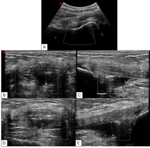
Supplementary Figure 1. Ultrasound view after stem cell injection into the intraarticular space of the (A) right hip joint, (B) right knee joint in sagittal view, (C) right knee joint in transversal view, (D) left knee joint in sagittal view, (E) left knee joint in transversal view.
Post-procedural evaluation showed persistent but non-worsening pain in the left hip, while left knee pain decreased significantly to 1 compared to baseline following the interventional pain management (IPM) sessions. The patient was admitted for one day for close observation of potential adverse reactions. Oral medications prescribed included diacerein every 12 hours, etoricoxib once daily, and eperisone every 12 hours. She was discharged after an uneventful observation period.
Weekly follow-up was conducted for the subsequent two months. Serial ultrasound monitoring demonstrated progressive reduction of intra-articular effusion in both the hip and knee joints, with complete resolution of effusion in the left knee during the later visits, as summarized in Table 1. Pain levels remained stable, and repeat stem cell injections at the same dosage were administered to the same anatomical sites. Thereafter, follow-up visits were scheduled every two weeks for nine months up to the present. At the latest evaluation, the patient reported being pain-free and able to perform daily activities without limitation or additional complaints.
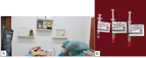
Supplementary Figure 2. (A) Ultrasound-guided interventional pain management. (B) Stem cell products used in the procedure
Osteoarthritis (OA) is a leading cause of disability in the elderly population, most commonly affecting the knee followed by the hip. The lifetime risk of developing symptomatic hip OA by the age of 85 is estimated at 25%. Intrinsic risk factors include age, sex, and genetic predisposition, while extrinsic factors involve repetitive mechanical stress from daily activities, increased body mass index, high-intensity sports, or physically demanding occupations. These factors may lead to repetitive microtrauma and structural joint damage.1 Histopathologically, articular cartilage has limited regenerative capacity due to its avascular nature, resulting in progressive chondrocyte loss, synovial hypertrophy, and subchondral bone changes.3 Mesenchymal stem cells (MSCs) have emerged as promising regenerative therapies due to their chondrogenic differentiation potential and paracrine anti-inflammatory and immunomodulatory effects.1-3
Several studies have shown promising results. Carlo Dall’Oca reported six patients with hip OA who received intra-articular injections of autologous adipose-derived MSCs, demonstrating significant improvement in Harris Hip Score (HHS) and reduction in WOMAC scores after six months, without significant adverse effects.5 In knee OA, several clinical trials have evaluated the role of MSCs. A study by Aryana compared secretome injections with hyaluronic acid, showing that HA provided better short-term pain reduction than secretome. However, other studies have demonstrated that MSCs can improve joint function and reduce pain for at least six months, although level 1 evidence for hip OA remains limited.6
Use of MSCs in hip osteoarthritis has been rarely reported, as most studies focus on knee OA. Yunong et al. demonstrated significant improvements in pain and function following intra-articular UC-MSC injections, with mostly transient adverse events.7 Similarly, a single UC-MSC injection showed superior improvements in WOMAC, pain scores, and quality of life compared with triamcinolone without serious complications.8 Moniruzzaman (2017) conducted a study involving 6 patients with OA ranging from Kellgren-Lawrence 3-4 on both knees. The patients all underwent intraarticular injections of bone marrow aspirate concentrate MSCs (BM-MSCs) and followed up for 6 months. These procedures successfully resulted in decreased tibiofemoral degeneration and increased cartilage thickness, resulting in increased joint space width, showing signs towards improvement of Kellgren-Lawrence grading of knee OA.9
In this case, umbilical cord–derived mesenchymal stem cells (UC-MSCs) were selected due to their higher proliferative capacity, stronger paracrine anti-inflammatory effects, and greater potential for cartilage regeneration compared with other stem cell sources. Several studies have compared UC-MSCs and bone marrow aspirate concentrate (BMAC) in the treatment of osteoarthritis. A case review involving 176 patients reported that both BMAC and UC-MSC therapies improved pain, functional outcomes, and quality of life; however, UC-MSC implantation demonstrated superior cartilage repair compared with BMAC.10 Similarly, Lee et al. found that although both therapies improved clinical outcomes in patients with medial unicompartmental knee OA undergoing high tibial osteotomy, UC-MSC treatment resulted in more effective cartilage regeneration.11 A systematic review and meta-analysis by Park et al. also reported comparable clinical outcomes between the two treatments, but superior cartilage repair with UC-MSCs based on International Cartilage Repair Society Cartilage Repair Assessment grades.12
In this case report, the patient received intra-articular stem cell injections in both the hip and knee joints. Clinically, the outcomes demonstrated symptomatic improvement, particularly in the left knee, although persistent pain in the left hip was still reported. These findings are consistent with the literature, which suggests that regenerative therapy using MSCs may provide analgesic and anti-inflammatory effects and improve joint function. However, the long-term benefits and potential structural regeneration remain subjects of ongoing investigation.
Stem cell injections may cause side effects similar to other intra-articular injection therapies, including joint swelling, post-procedural contusion or hematoma, injection site pain, and mild joint effusion. Fortunately, in our patient, the only post-procedural concern was persistent pain without change in the NRS score within the first 24 hours after injection. The pain subsequently subsided, and the patient is currently pain-free. Another important risk of stem cell therapy is the potential for oncogenic transformation, which may arise from mutation accumulation, clonal expansion of pre-existing or culture-acquired driver mutations, and prolonged stem cell lifespan following transplantation. For this reason, baseline tumor marker evaluation was performed prior to stem cell administration, and accurate ultrasound-guided delivery was used to ensure precise intra-articular injection.13
Ultrasound-guided intra-articular injection has consistently demonstrated superior performance compared with landmark-guided techniques. Previous studies have shown that ultrasound guidance improves injection accuracy across multiple joints, particularly in deeper joints such as the hip.14 A recent meta-analysis also reported improved clinical outcomes with ultrasound-guided injections, including reduced procedural pain, greater pain reduction, improved function, and higher patient satisfaction in knee osteoarthritis.15 Together, these findings suggest that ultrasound guidance enhances both technical precision and patient-reported outcomes, supporting its superiority over blind intra-articular injection techniques.
Regenerative therapies such as stem cell treatment represent a promising alternative for patients with hip and knee osteoarthritis who do not respond adequately to conservative management. In this case, clinical improvement was achieved through careful patient selection, appropriate laboratory screening, and precise ultrasound-guided intra-articular injection. However, potential risks, including post-injection reactions and the theoretical risk of oncogenic transformation, highlight the importance of thorough pre-procedural evaluation and accurate injection techniques. A limitation of this report is the absence of follow-up radiologic assessment. Future studies incorporating serial imaging and long-term follow-up are necessary to better determine the safety, efficacy, and potential disease-modifying effects of mesenchymal stem cell therapy in osteoarthritis.
5. Ethics Approval of Research
This case report was conducted in accordance with the ethical principles outlined in the Declaration of Helsinki. Written informed consent was obtained from the patient for participation in the treatment and for the publication of clinical data and images in this report. Institutional ethical approval was obtained from Ethics Committee of the Faculty of Medicine, Padjadjaran University, Bandung (number 732/UN6.KEP/EC/2022).
6. Acknowledgement
The authors would like to thank the medical staff of Bandung Pain Rehab Center for their assistance in the clinical management of the patient and data collection for this case report.
7. Author Contributions
TCNT conceived and supervised the study, performed the ultrasound-guided procedures, and was responsible for clinical management and final manuscript revision. CL contributed to clinical data acquisition. DGSP provided orthopedic consultation and manuscript review. MJY and SDZ contributed to data collection, literature review, and manuscript drafting. DH and YY contributed to patient management, data interpretation, and manuscript review. All authors read and approved the final version of the manuscript.
8. Conflict of Interest
The authors declare that there are no potential conflicts of interest to declare.
Authors affiliation:
- Theresia Chandra Tania Novy, Bandung Pain Rehab Center, Bandung 40115, West Java, Indonesia; E-mail: theresianovymd@gmail.com Phone: +62 811 2246 067
- Christopher Lampah. Physical Medicine & Rehabilitation Program, Universitas Sam Ratulangi, Bahu, Manado 95115, North Sulawesi, Indonesia; Email: chritopherlampah@gmail.com
- Dewa Gede Satria Ambara Putra, Orthopedic Department of Balimed Hospital, Denpasar 80117, Bali, Indonesia; Email: satriaortopedi22@gmail.com
- Daniel Hadimartana, PIK Pain Rehab Center, Jakarta 14470, Indonesia; Email: danielhadimartana@gmail.com
- Maria Jessica Yaputri, Bandung Pain Rehab Center, Bandung 40115, West Java, Indonesia; Email: mjessicayaputri@gmail.com
ABSTRACT
Introduction: Osteoarthritis (OA) is a progressive degenerative joint disease commonly affecting weight-bearing joints such as the knee and hip, leading to pain, stiffness, and functional impairment. Conventional treatments often provide only temporary symptom relief, prompting increasing interest in regenerative approaches such as mesenchymal stem cell therapy.
Case Report: We report a 60-year-old woman with a one-year history of left hip and knee pain refractory to conservative management. Clinical and imaging findings were consistent with osteoarthritis and suspected avascular necrosis of the left femoral head. Initial ultrasound examination demonstrated intra-articular effusion in both joints. The patient first underwent ultrasound-guided intra-articular corticosteroid injection followed by viscosupplementation. Due to persistent symptoms, regenerative therapy using umbilical cord–derived mesenchymal stem cells (UC-MSCs) was performed under ultrasound guidance. A total of 70 × 10⁶ cells were injected intra-articularly, including 20 × 10⁶ cells into each knee and 30 × 10⁶ cells into the left hip. Progressive improvement in pain and function was observed without significant adverse events.
Conclusion: Ultrasound-guided intra-articular UC-MSC therapy may represent a promising minimally invasive treatment option for patients with osteoarthritis who do not respond adequately to conventional therapy.
Keywords: hip osteoarthritis; knee osteoarthritis; mesenchymal stem cells; regenerative therapy; ultrasound-guided injection
Citation: Novy TCT, Lampah C, Putra DGS, Hadimartana D, Yaputri MJ. Ultrasound-guided stem cell therapy for hip and knee osteoarthritis: a case report. Anaesth. pain intensive care 2026;30(6):773-779.
DOI: 10.35975/apic.v30i6.3274
Received: xxxxxxxxx; Revised: xxxxxxxxxxx; Accepted: xxxxxxxxxx
1. INTRODUCTION
Osteoarthritis (OA) is the most prevalent degenerative joint disorder, characterized by chondrocyte damage caused by inflammatory cytokines and mechanical stress, leading to joint space narrowing, osteophyte formation, and subchondral bone changes. Globally, approximately 240 million adults suffer from symptomatic OA, and its prevalence continues to increase with aging populations. The main symptoms include pain, stiffness, and limited joint motion. Risk factors include genetics, sex, mechanical stress, and previous trauma, with the knee most commonly affected, followed by the hip.1 Diagnosis is usually clinical, although radiographic grading systems such as Tönnis and Kellgren–Lawrence are widely used to assess disease severity.2 Current management includes weight reduction, physiotherapy, pharmacologic therapy, and intra-articular injections, while joint arthroplasty remains invasive and associated with potential complications.3 Mesenchymal stem cells (MSCs) have emerged as a promising regenerative therapy due to their immunomodulatory, anti-inflammatory, and cartilage-regenerative potential.⁴ This case report aims to present a regenerative intervention using umbilical-derived stem cells in a 60-year-old woman with hip and knee OA.
2. CASE PRESENTATION
We report a 60-year-old woman presenting with a one-year history of left buttock and hip pain with a Numeric Rating Scale (NRS) score of 8. The pain was aggravated by standing up and sitting down and was accompanied by mild left knee pain during weight-bearing activities such as standing and walking. The patient had previously consulted an orthopaedic specialist and a neurologist and received pharmacological therapy, but without significant improvement.
The pain later recurred, predominantly involving the left knee and thigh, particularly when rising from bed or changing position from sitting to standing. She also reported sleep disturbances caused by pain during positional changes. Her daily activities included running a shop and baking, which required prolonged standing, as well as exercising on a stationary bicycle.
Physical examination revealed an antalgic gait and tenderness over the right hip joint. Provocative low back maneuvers on the left side were positive, suggesting hip osteoarthritis, left piriformis syndrome, and left sacroiliitis. Examination of the left knee demonstrated joint crepitus and mild tenderness over the suprapatellar bursa, medial collateral ligament (MCL), and pes anserine bursa, suggesting bursitis and knee osteoarthritis as possible differential diagnoses.
Radiographic evaluation of both knees and the left hip joint is shown in Figure 1. Knee radiographs demonstrated osteophyte formation consistent with mild bilateral knee osteoarthritis (Figure 1A–B), while hip radiography revealed degenerative changes in the left hip joint (Figure 1C). Previous MRI findings further indicated suspected avascular necrosis (AVN) of the left femoral head with secondary osteoarthritis of the left hip and a superior labral tear. A cystic lesion containing both fat and solid components was also observed in the left pelvic cavity. MRI images of the pelvis and hip region are presented in Figure 1D–E.
Figure 1. Radiographic and MRI findings of the knee and hip joints. (A) Anteroposterior radiograph of the right knee demonstrating osteophyte formation consistent with mild osteoarthritis. (B) Anteroposterior radiograph of the left knee showing similar osteoarthritic changes. (C) Anteroposterior radiograph of the left hip revealing degenerative changes suggestive of secondary osteoarthritis. (D–E) Magnetic resonance imaging (MRI) of the pelvis and hip region demonstrating suspected avascular necrosis (AVN) of the left femoral head with associated secondary osteoarthritis and a superior labral tear. A cystic lesion containing both fat and solid components is also observed within the left pelvic cavity.
At the initial visit, ultrasound examination demonstrated joint effusion and osteophytes in the hip and knee joints. Quantitative measurements of intra-articular effusion are summarized in Table 1, which showed greater effusion in the left hip joint and mild effusion in both knees. Ultrasound-guided intra-articular injection with triamcinolone was subsequently performed as part of interventional pain management (IPM), and the patient was prescribed oral medications including nonsteroidal anti-inflammatory drugs (NSAIDs), a muscle relaxant, and vitamins.
Two weeks later, the patient was re-evaluated and her NRS score had decreased to 3, indicating mild pain. Physical examination demonstrated resolution of the antalgic gait with marked reduction in tenderness and pain during provocative maneuvers and range-of-motion testing of both the hip and knee. Follow-up ultrasound showed reduced effusion in the left hip joint and minimal residual effusion in both knees compared with the initial evaluation, as summarized in Table 1. During this visit, ultrasound-guided viscosupplementation of the left hip and both knees was performed.
| Table 1. Effusion measurement of the intraarticular space | ||||||
| Effusion measurement (cm) | Hip Joint | Knee Joint | ||||
| Right | Left | Right (longitudinal view) | Left (longitudinal view) | Right (sagittal view) | Left (sagittal view) | |
| First visit | 0.62 | 0.87 | 0.45 | 0.36 | 0.37 | 0.24 |
| Second visit | 0.59 | 0.63 | 0.33 | 0.27 | 0.14 | 0.15 |
| Third visit | 0.62 | 0.67 | 0.45 | 0.35 | 0.21 | 0.23 |
| Fourth visit | 0.55 | 0.59 | 0.11 | No effusion | 0.13 | No effusion |
The patient was subsequently scheduled for regenerative therapy using stem cell injection for hip and knee osteoarthritis. Two weeks later, she reported minimal pain with a stable NRS score of 3. Laboratory investigations were within normal limits. These tests were performed as a precaution prior to stem cell therapy due to potential risks including carcinogenicity and immunologic reactions. Evaluations included complete blood count, total IgE levels, platelet aggregation testing, and tumor markers including CEA, AFP, CA 19-9, CA 125, and CA 15-3 to exclude occult malignancy and establish a baseline prior to treatment.
After confirming that all laboratory results were within normal limits, a repeat physical examination was performed, yielding findings similar to those of the previous visit. The procedure was carried out using ultrasound-guided interventional pain management (IPM), as illustrated in Supplementary Figure 2A, with the stem cell product shown in Supplementary Figure 2B. Stem cell therapy was administered using umbilical cord–derived mesenchymal stem cells (UC-MSCs) supplied by PT Bifarma Adiluhung, with 1 mL diluted in 0.9% NaCl. A total dose of 70 × 10⁶ cells was administered, consisting of 20 × 10⁶ cells injected into the right knee joint, 20 × 10⁶ cells into the left knee joint, and 30 million cells into the left hip joint. Accurate intra-articular delivery was ensured under real-time ultrasound visualization. Post-injection ultrasound images demonstrating the intra-articular space of the hip and knee joints are presented in Supplementary Figure 1.
Supplementary Figure 1. Ultrasound view after stem cell injection into the intraarticular space of the (A) right hip joint, (B) right knee joint in sagittal view, (C) right knee joint in transversal view, (D) left knee joint in sagittal view, (E) left knee joint in transversal view.
Post-procedural evaluation showed persistent but non-worsening pain in the left hip, while left knee pain decreased significantly to 1 compared to baseline following the interventional pain management (IPM) sessions. The patient was admitted for one day for close observation of potential adverse reactions. Oral medications prescribed included diacerein every 12 hours, etoricoxib once daily, and eperisone every 12 hours. She was discharged after an uneventful observation period.
Weekly follow-up was conducted for the subsequent two months. Serial ultrasound monitoring demonstrated progressive reduction of intra-articular effusion in both the hip and knee joints, with complete resolution of effusion in the left knee during the later visits, as summarized in Table 1. Pain levels remained stable, and repeat stem cell injections at the same dosage were administered to the same anatomical sites. Thereafter, follow-up visits were scheduled every two weeks for nine months up to the present. At the latest evaluation, the patient reported being pain-free and able to perform daily activities without limitation or additional complaints.
Supplementary Figure 2. (A) Ultrasound-guided interventional pain management. (B) Stem cell products used in the procedure
3. DISCUSSION
Osteoarthritis (OA) is a leading cause of disability in the elderly population, most commonly affecting the knee followed by the hip. The lifetime risk of developing symptomatic hip OA by the age of 85 is estimated at 25%. Intrinsic risk factors include age, sex, and genetic predisposition, while extrinsic factors involve repetitive mechanical stress from daily activities, increased body mass index, high-intensity sports, or physically demanding occupations. These factors may lead to repetitive microtrauma and structural joint damage.1 Histopathologically, articular cartilage has limited regenerative capacity due to its avascular nature, resulting in progressive chondrocyte loss, synovial hypertrophy, and subchondral bone changes.3 Mesenchymal stem cells (MSCs) have emerged as promising regenerative therapies due to their chondrogenic differentiation potential and paracrine anti-inflammatory and immunomodulatory effects.1-3
Several studies have shown promising results. Carlo Dall’Oca reported six patients with hip OA who received intra-articular injections of autologous adipose-derived MSCs, demonstrating significant improvement in Harris Hip Score (HHS) and reduction in WOMAC scores after six months, without significant adverse effects.5 In knee OA, several clinical trials have evaluated the role of MSCs. A study by Aryana compared secretome injections with hyaluronic acid, showing that HA provided better short-term pain reduction than secretome. However, other studies have demonstrated that MSCs can improve joint function and reduce pain for at least six months, although level 1 evidence for hip OA remains limited.6
Use of MSCs in hip osteoarthritis has been rarely reported, as most studies focus on knee OA. Yunong et al. demonstrated significant improvements in pain and function following intra-articular UC-MSC injections, with mostly transient adverse events.7 Similarly, a single UC-MSC injection showed superior improvements in WOMAC, pain scores, and quality of life compared with triamcinolone without serious complications.8 Moniruzzaman (2017) conducted a study involving 6 patients with OA ranging from Kellgren-Lawrence 3-4 on both knees. The patients all underwent intraarticular injections of bone marrow aspirate concentrate MSCs (BM-MSCs) and followed up for 6 months. These procedures successfully resulted in decreased tibiofemoral degeneration and increased cartilage thickness, resulting in increased joint space width, showing signs towards improvement of Kellgren-Lawrence grading of knee OA.9
In this case, umbilical cord–derived mesenchymal stem cells (UC-MSCs) were selected due to their higher proliferative capacity, stronger paracrine anti-inflammatory effects, and greater potential for cartilage regeneration compared with other stem cell sources. Several studies have compared UC-MSCs and bone marrow aspirate concentrate (BMAC) in the treatment of osteoarthritis. A case review involving 176 patients reported that both BMAC and UC-MSC therapies improved pain, functional outcomes, and quality of life; however, UC-MSC implantation demonstrated superior cartilage repair compared with BMAC.10 Similarly, Lee et al. found that although both therapies improved clinical outcomes in patients with medial unicompartmental knee OA undergoing high tibial osteotomy, UC-MSC treatment resulted in more effective cartilage regeneration.11 A systematic review and meta-analysis by Park et al. also reported comparable clinical outcomes between the two treatments, but superior cartilage repair with UC-MSCs based on International Cartilage Repair Society Cartilage Repair Assessment grades.12
In this case report, the patient received intra-articular stem cell injections in both the hip and knee joints. Clinically, the outcomes demonstrated symptomatic improvement, particularly in the left knee, although persistent pain in the left hip was still reported. These findings are consistent with the literature, which suggests that regenerative therapy using MSCs may provide analgesic and anti-inflammatory effects and improve joint function. However, the long-term benefits and potential structural regeneration remain subjects of ongoing investigation.
Stem cell injections may cause side effects similar to other intra-articular injection therapies, including joint swelling, post-procedural contusion or hematoma, injection site pain, and mild joint effusion. Fortunately, in our patient, the only post-procedural concern was persistent pain without change in the NRS score within the first 24 hours after injection. The pain subsequently subsided, and the patient is currently pain-free. Another important risk of stem cell therapy is the potential for oncogenic transformation, which may arise from mutation accumulation, clonal expansion of pre-existing or culture-acquired driver mutations, and prolonged stem cell lifespan following transplantation. For this reason, baseline tumor marker evaluation was performed prior to stem cell administration, and accurate ultrasound-guided delivery was used to ensure precise intra-articular injection.13
Ultrasound-guided intra-articular injection has consistently demonstrated superior performance compared with landmark-guided techniques. Previous studies have shown that ultrasound guidance improves injection accuracy across multiple joints, particularly in deeper joints such as the hip.14 A recent meta-analysis also reported improved clinical outcomes with ultrasound-guided injections, including reduced procedural pain, greater pain reduction, improved function, and higher patient satisfaction in knee osteoarthritis.15 Together, these findings suggest that ultrasound guidance enhances both technical precision and patient-reported outcomes, supporting its superiority over blind intra-articular injection techniques.
4. CONCLUSION
Regenerative therapies such as stem cell treatment represent a promising alternative for patients with hip and knee osteoarthritis who do not respond adequately to conservative management. In this case, clinical improvement was achieved through careful patient selection, appropriate laboratory screening, and precise ultrasound-guided intra-articular injection. However, potential risks, including post-injection reactions and the theoretical risk of oncogenic transformation, highlight the importance of thorough pre-procedural evaluation and accurate injection techniques. A limitation of this report is the absence of follow-up radiologic assessment. Future studies incorporating serial imaging and long-term follow-up are necessary to better determine the safety, efficacy, and potential disease-modifying effects of mesenchymal stem cell therapy in osteoarthritis.
5. Ethics Approval of Research
This case report was conducted in accordance with the ethical principles outlined in the Declaration of Helsinki. Written informed consent was obtained from the patient for participation in the treatment and for the publication of clinical data and images in this report. Institutional ethical approval was obtained from Ethics Committee of the Faculty of Medicine, Padjadjaran University, Bandung (number 732/UN6.KEP/EC/2022).
6. Acknowledgement
The authors would like to thank the medical staff of Bandung Pain Rehab Center for their assistance in the clinical management of the patient and data collection for this case report.
7. Author Contributions
TCNT conceived and supervised the study, performed the ultrasound-guided procedures, and was responsible for clinical management and final manuscript revision. CL contributed to clinical data acquisition. DGSP provided orthopedic consultation and manuscript review. MJY and SDZ contributed to data collection, literature review, and manuscript drafting. DH and YY contributed to patient management, data interpretation, and manuscript review. All authors read and approved the final version of the manuscript.
8. Conflict of Interest
The authors declare that there are no potential conflicts of interest to declare.
9. REFERENCES
- Mardones R, Jofré CM, Tobar L, Minguell JJ. Mesenchymal stem cell therapy in the treatment of hip osteoarthritis. J Hip Preserv Surg 2017;4:159-63.
- Pracheta Ewari GA, Aryana IGNW. The use of mesenchymal stem cell secretome in osteoarthritis: a literature review. Int J Res Rev 2024;11:359-70.
- Gibbs AJ, Gray B, Wallis JA, Taylor NF, Kemp JL, Hunter DJ, et al. Recommendations for the management of hip and knee osteoarthritis: a systematic review of clinical practice guidelines. Osteoarthritis Cartilage 2023;31:1280-92.
- Giorgino R, Alessandri Bonetti M, Migliorini F, Nannini A, Vaienti L, Peretti GM, et al. Management of hip osteoarthritis: harnessing the potential of mesenchymal stem cells—a systematic review. Eur J Orthop Surg Traumatol 2024;34:3847-57.
- Dall’Oca C, Breda S, Elena N, Valentini R, Samaila EM, Magnan B. Mesenchymal stem cells injection in hip osteoarthritis: preliminary results. Acta Biomed 2019;90:75-80.
- Aryana IGNW, Suyasa IK, Dharmayuda CGO, Gunawan D. Clinical outcomes comparison in administration of secretome vs hyaluronic acid in patients with knee osteoarthritis Kellgren-Lawrence grade I-III. Indones J Med 2024;9:467-77.
- Ao Y, Duan J, Xiong N, Qian N, Zhang R, Yang L, et al. Repeated intra-articular injections of umbilical cord-derived mesenchymal stem cells for knee osteoarthritis: a phase I, single-arm study. BMC Musculoskelet Disord 2023;24:1021.
- Pico OA, Espinoza F, Cádiz MI, Sossa CL, Becerra-Bayona SM, Salgado MCC, et al. Efficacy of a single dose of cryopreserved human umbilical cord mesenchymal stromal cells for the treatment of knee osteoarthritis: a randomized, controlled, double-blind pilot study. Cytotherapy 2025;27:188-200.
- Moniruzzaman M. Bone marrow aspirate concentrate mesenchymal stem cells in osteoarthritis knee: case series. Presented at: drZaman’s Interventional Pain, Arthritis, Spine & Stem Cell Center; 2017; Dhaka, Bangladesh.
- Zhang Y, Zhuang H, Ren X, Jiang F, Zhou P. Therapeutic effects of different intervention forms of human umbilical cord mesenchymal stem cells in the treatment of osteoarthritis. Front Cell Dev Biol 2023;11:1246504.
- Lee NH, Na SM, Ahn HW, Kang JK, Seon JK, Song EK. Allogenic human umbilical cord blood-derived mesenchymal stem cells are more effective than bone marrow aspiration concentrate for cartilage regeneration after high tibial osteotomy in medial unicompartmental osteoarthritis of the knee. Arthroscopy 2021;37:2521-30.
- Park D, Choi YH, Kang SH, Koh HS, In Y. Bone marrow aspirate concentrate versus human umbilical cord blood-derived mesenchymal stem cells for combined cartilage regeneration procedure in patients undergoing high tibial osteotomy: a systematic review and meta-analysis. Medicina (Kaunas) 2023;59:634.
- Fathani IJ, Budiyanto MHA, Jannatin N. Therapeutic modality of mesenchymal stem cells for moderate to severe knee osteoarthritis: a systematic review. JOINTS 2025;14:46-55.
- Saha P, Smith M, Hasan K. Accuracy of intraarticular injections: blind vs image guided techniques—a review of literature. J Funct Morphol Kinesiol 2023;8:93.
- Oo WM, Linklater J, Siddiq MAB, Fu K, Hunter DJ. Comparison of ultrasound guidance with landmark guidance for symptomatic benefits in knee, hip and hand osteoarthritis: systematic review and meta-analysis of randomised controlled trials. Australas J Ultrasound Med 2024;27:97-105.