
Mert Akbas1, Arif Yegin2, Huseyin Yilmaz3, R. Emre Altekin4, Muhammed Jamil Sabit5, Suat Sanli1
1Associate Professor; 2Professor; 5Consultant Algologist
Department of Anesthesiology, Division of Algology, Akdeniz University Medical Faculty, Antalya, (Turkey)
3Professor; 4Assistant Professor
Department of Cardiology, Akdeniz University Medical Faculty, Antalya, (Turkey)
Correspondence: Mert Akbas, MD, Akdeniz University Medical Faculty, Department of Anesthesiology, Antalya 07070, (Turkey); Tel: +90.242.2496642; Fax: +90.242.2415426; E-mail akbasmert@akdeniz.edu.tr
ABSTRACT
Angina pectoris is characterized by an oppressive pain in the chest due to inadequate blood flow to the heart muscle. If patients with chronic angina are refractory to conservative therapies and cannot undergo coronary revascularization surgery, spinal cord stimulation can be proposed as a possible treatment option. We present a case of a 70 years old female patient, who presented with angina pectoris at rest, which could not be relieved although surgical revascularization and cardiac stenting were performed previously. Angiography was performed but no lesions were found to require the repetition of surgery or cardiac stenting. She could only walk up to 20 meters despite maximum medical therapies. During sleep, chest pain woke her up. An eight-contact paddle electrode was placed with its cephalad tip at the T1–T4 level on the left of the midline followed by test stimulations. The test stimulations should extend over the areas that are painful during angina attacks. The symptoms of the patient improved remarkably in 24 hours. His nitrate consumption could be reduced by 80% by the end of 3 months. If all available medical and invasive treatment options fail, SCS can be used as a viable alternative for treatment.
Key words: Spinal cord stimulation, Electric Stimulation Therapy/methods; Angina Pectoris/therapy; Electrical Equipment and Supplies; Electrode; Quality of Life
Citation: Akbas M, Yegin A, Yilmaz H, Altekin RE, Sabit MJ, Sanli S. Unstable angina pectoris and spinal cord stimulation. Anaesth Pain & Intensive Care 2015;19(3):383-385
INTRODUCTION
Angina pectoris is characterized by an oppressive pain in the chest due to inadequate blood flow to the heart muscle. Vasodilators can be administered or exertion can be reduced for the treatment of angina pectoris. There are a lot of pharmacological therapies available today. Moreover, in case of a finding that indicates a single or more than one discrete area that is obstructed, angina attacks can be relieved through revascularization procedures such as percutaneous coronary angioplasty and coronary bypass surgery (CBS). One or more percutaneous coronary angioplasties or even CBS are often performed for these patients besides extensive anti-angina pharmacological treatment.
If patients with chronic angina are refractory to conservative therapies and cannot undergo coronary revascularization surgery, spinal cord stimulation (SCS) can be proposed as a possible treatment option.1
This case report presents the application of SCS in a 70-year-old woman who suffered from symptomatic ischemic heart disease for a decade and who did not respond to all available treatment options for intractable severe chest pain. Furthermore, this represents the first percutaneous application of spinal cord stimulator for intractable angina pectoris in Turkey.
CASE REPORT
The patient who presented 15 years ago with angina pectoris (stable angina), underwent coronary angiography, and was advised to have coronary artery by-pass grafting (CABG) due to 95% stenosis in the proximal part of her left anterior descending artery (LAD), 60% stenosis in the proximal part of right coronary artery (RCA) and 60% stenosis in the mid portion and obtuse margin of circumflex artery (CX). Then left internal mammary artery (LIMA)-LAD by-pass was performed for the patient.
The coronary angiography performed due to the relapsing chest pain 2 years after the operation revealed the bypass graft patency, 80% stenosis in RCA and 80% stenosis in CX as well as severe lesions at other sites. Bare metal stents were implanted into both lesions. After the procedure the patient relieved, but soon had recurrence of repeated pains. Therefore, another coronary angiography was performed and it showed restenosis in the CX stent despite the patency of RCA and by-pass grafts. For that restenosis, brachy therapy (a procedure that involves placing radioactive material inside body) was decided. Brachytherapy was applied to that stenotic lesion while kissing percutaneous transluminal coronary angioplasty (PTCA) + bare metal stent was implanted to the LAD-CX lesion. As there were critical lesions below and above the RCA stent too, that was implanted before, a bare metal stent was implanted for each location.
However, the patient suffered from repeat chest pain again in the same year. The angiography performed demonstrated that a patent by-pass graft-CX, but restenosis in LDA and RCA stents. An additional revascularization was considered to be risky for the patient; therefore, the cardiologists decided to apply intensive medical therapy, which included metoprolol, nifedipine, isosorbide mononitrate, candesartan cilexetil, acetyl salicylic acid, atorvastatin, clopidogrel and ivabradine.
The patient had to be hospitalized occasionally due to episodes of relapsing chest pain despite the medical therapy. Left stellate ganglion block was also tried for her refractory angina with temporary relief only. Ultimately spinal cord stimulation was proposed and she was referred to our outpatient algology service.
An informed consent was obtained from the patient, 18G cannula was inserted into a peripheral vein. Normal saline was infused intravenously @5 mL/kg/hr. A 20G radial arterial catheter was inserted into the left hand for continuous blood pressure monitoring. In the operating room, patient was continuously monitored throughout the procedure via ECG, invasive blood pressure (IBP), heart rate (HR), and peripheral oxygen saturation (SpO2). Supplemental oxygen was given via a nasal cannula. Sedation was induced with 1.0 mg midazolam and 0.05 mg fentanyl IV, and repeated when needed. The patient was monitored in the recovery unit until she was discharged when her memory, calculation, and orientation were normalized. No adverse events were observed.
The operation was performed under local anesthesia to allow the patient to answer questions during the intra operative stimulation. The epidural space was punctured at the level of L1-L2. The neuro stimulation lead Lamitrode™ (St. Jude Medical) was advanced through the epidural space and the tip was positioned in the midline or a few millimetres to the left at the T1-T4 level (Figures 1 & 2) so that the patient could feel a prickling sensation in the precordial area and into the arms.
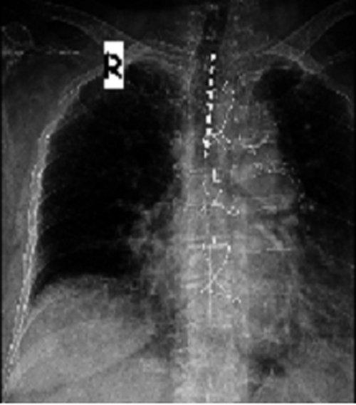
Figure 1: AP view of T1-T4 lead
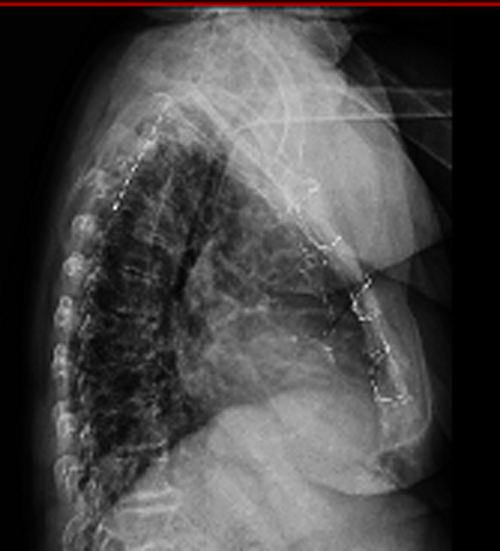
Lateral view, T1-T4 lead (posterior)
The distal end of the electrode was sutured to the fascia and connected via a tunnelled extension lead to the external pulse generator. The pulse width was 200 microseconds and the frequency was 80 Hz. Appropriate amplitude (usually 8-10 V) was used for comfortable paraesthesia. The study consisted of two parts: trial period (2 weeks) to standardise the stimulation when mobilization was performed. After a successful trial period the generator was implanted.
The stimulation was carried out for 30 min 10-12 times a day during the trial period and five to six times a day during the treatment period. Altogether the heart rate and systolic blood pressure were lowered slightly. VAS and nitrate consumption was reduced by up to 80%.
DISCUSSION
Refractory angina pectoris is a medical condition in which angina pain cannot be relieved through either anti-angina medication or revascularization procedures.1,2 According to the findings of randomized studies on SCS, angina complaints can be reduced, use of short-acting nitrates decreased, and an improvement in quality of life achieved in addition to the increased exercise capacity. 80% of patients feel the beneficial effects for at least 1 year, while 60% of patients have reported that their exercise capacity increased and quality of life improved for up to 5 years.3
Anti-angina effects help reduce ischemia, which is shown by exercise stress test and ambulatory ECG monitoring. Chauhan reported that coronary flow velocity increased during neuromodulation.3 Myocardial oxygen consumption is decreased due to a possible redistribution of the blood flow to the ischemic tissue.4-6
The mechanism of action of SCS still remains unclear. Some mechanisms proposed have been summarized in a recent review of experimental studies.7 Increased blood flow is not possibly the only mechanism that provides benefits to the heart. It has been recently demonstrated that my ocardial ischemia can be improved through redistribution of local blood flow.8 The intrinsic cardiac nervous system can also be normalized by SCS.9
Infection, lead migration, and device failure are found to be the most common complications in the studies reviewed. Each of these complications developed in less than one percent of the participants, with no permanent sequelae recorded. In our patient we did not observe any complication. In the long term follow up period, the patient did not report any severe angina pain, while her nitroglycerine consumption and verbal numerical rating score declined approximately by 80%. Also her sleep quality and daily exercise capacity improved.
REFERENCES
- van Kleef M, Staats P, Mekhail N, Huygen F. Chronic refractory angina pectoris. Pain Pract. 2011 Sep-Oct;11(5):476-82. Review.[PubMed] doi: 10.1111/j.1533-2500.2010.00444.x.
- Eliasson T, Augustinsson LE, Mannheimer C. Spinal Cord Stimulation in severe angina pectoris—presentation of current studies, indications and clinical experience. Pain 1996; 65:169-179. [PubMed]
- Deer TR, Raso LJ. Spinal cord stimulation for refractory angina pectoris and peripheral vascular disease. Pain Physician. 2006 Oct;9(4):347-52.[PubMed][Free full text]
- Mannheimer C, Eliasson T, Andersson B, Bergh CH, Augustinsson LE, Emanuelsson H, Waagstein F. Effects of spinal cord stimulation in angina pectoris induced by pacing and possible mechanisms of action. BMJ 1993; 307:477- 480. [PubMed] [Free full text]
- Hautvast RW, DeJongste MJ, ter Horst FJ, Blanksma PK, Lie KI. Angina pectoris refractory for conventional therapy—is neurostimulation a possible alternative treatment? Clin Cardiol 1996; 19:531-5.[PubMed]
- Mobilia G, Zuin G, Zanco P, Di Pede F, Pinato G, Neri G, et al. Effects of spinal cord stimulation on regional myocardial blood flow in patients with refractory angina. A positron emission tomography study. G Ital Cardiol 1998; 28:1113-9.[PubMed]
- Ekre O, Eliasson T, Norrsell H, Währborg P, Mannheimer C: Electrical Stimulation versus Coronary Artery Bypass Surgery in Severe Angina Pectoris. Long-term effects of spinal cord stimulation and coronary artery bypass grafting on quality of life and survival in the ESBY study. Eur Heart J 2002, 23(24):1938-45.[PubMed]
- De Vries J, Foreman RD, DeJongste MJ: The anti-ischemic effects of electrical neuro stimulation in the heart. Cleve Clin J Med 2007, 74(Suppl 1):S42-7. [PubMed]
- Wu M, Linderoth B, Foreman RD: Putative mechanisms behind effects of spinal cord stimulation on vascular diseases: A review of experimental studies. Auton Neurosci 2008, 138:9-23. [PubMed][Free full text]
