
Sukhyanti Kerai, Lalit Sehrawat
Department of Anesthesiology & Critical Care, Maulana Azad Medical College & associated hospitals, New Delhi, (India).
Correspondence: Dr. S. Kerai, Department of Anesthesiology & Critical Care, Maulana Azad Medical College & Associated Hospitals, Bahadur Shah Zaffar Marg, New Delhi, (India); E-mail: drsukhi25@gmail.com
ABSTRACT
Mucopolysaccharidosis is a rare inherited lysosomal storage disease characterized by increasing deposition of unmetabolized glucosaminoglycans. The surgical procedures under general anesthesia in these patients is associated with high mortality due to difficulty in airway management .The regional anesthesia is useful alternative; however, previous case reports describing epidural/spinal anesthesia have reported failure of this technique. We describe a case of mucopolysaccharidosis type I (Hurler syndrome) for repair of umbilical and inguinal hernia repair conducted under epidural anesthesia with sedation, obviating the need of intubation.
Key words: Mucopolysaccharidosis; Hurler syndrome; Epidural anesthesia; Hernia repair
Citation: Kerai S, Sehrawat L. Successful epidural anesthesia in a patient of Hurler syndrome for hernia repair. Anaesth Pain & Intensive Care 2016;20(3):350-352
Received: 17 April 2016; Reviewed: 20 June 2016; Accepted: 20 June 2016
INTRODUCTION
Mucopolysaccharidosis (MPS) is a rare inherited lysosomal storage diseases associated with progressive accumulation of glucosaminoglycans (GAGs) in tissues and organs. Surgery and anesthesia in these patients are associated with high mortality.1 The most serious anesthetic complications are associated with airway obstruction, with accompanying difficulty in ventilation and oxygenation, resulting in significant cardiovascular compromise. We describe a case of mucopolysaccharidosis type I (Hurler syndrome) for repair of umbilical and inguinal hernia repair conducted under epidural anesthesia with sedation, obviating the need of intubation.
CASE REPORT
A 4 year old, 12 kg, boy presented for umbilical and bilateral inguinal hernia repair (Figure 1). A known case of Hurler syndrome, he had a history of snoring, delayed developmental milestones and mental retardation.

Figure 1: Child of Hurler syndrome with epigastric hernia and bilateral inguinal hernias
The airway examination revealed adequate mouth opening, large tongue, modified Mallampati class 4, short neck and adequate range of neck movements. His intellectual functions were below his age. Rest of neurological and other systemic examination was normal. No abnormalities were found on routine laboratory investigations and echocardiogram. X-ray spine showed low thoracic and lumbar kyphosis (Figure 2) and cervical lordosis. Atlantoaxial instability was not evident in cervical spine x-ray. Apart from usual preoperative advice, high risk consent was obtained from parents in view of an anticipated difficult airway (DA) and thus, preoperative sedative medications were avoided.
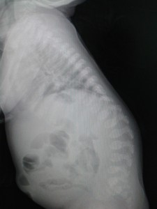
Figure 2: X-ray of lumbar spines showing kyphosis in lumbar spine
In operating room a difficult airway cart was prepared and senior anesthesiologists were present for case management. The patient was shifted to operating room accompanied by his mother to allay anxiety .The standard anesthetic monitors were attached and baseline values were noted. An intravenous cannula of 22 G was secured over dorsum of left hand which was pre-treated with EMLA cream. He was sedated slowly using infusion of dexmedetomidine starting with a bolus of 1 μg/kg over ten minutes, followed by 0.7 μg/kg/hr. The oxygen was supplemented using a facemask at 4 lit/min. Left lateral position was made and under all aseptic precautions epidural space was located at L2-3 interspace at depth of 3 cm using loss of resistance technique with air. The catheter was inserted 4 cm inside epidural space and fixed to the skin. The airway patency under sedation was constantly monitored by pattern of breathing and pulse oximetry. Through epidural catheter 5 ml of 0.5% bupivacaine was given slowly to achieve sensory block of T6 as assessed by pinprick. Surgery was started and the infusion of dexmedetomidine was continuously administered at 0.5-0.7 μg/kg/hr for sedation. After 45 min epidural blockade was supplemented further with 2 ml of 0.5% bupivacaine. No adverse hemodynamic events or episodes of airway obstruction were noted intraoperatively. The SpO2 remained within 94-98%. The surgical procedure was completed in 60 min. At the conclusion of surgery dexmedetomidine infusion was switched off and the patient regained consciousness within 20 min. Postoperatively he was shifted to postsurgical unit and analgesia was maintained through epidural catheter for two days with 0.125% bupivacaine along with intravenous paracetamol.
DISCUSSION
The airway management and endotracheal intubation in patients with Hurler syndrome is most challenging of all difficult airways. It is considered to be “the worst airway management problem a pediatric anesthesiologist could deal with.2 These patients develops progressive generalized infiltration and thickening of the soft tissues due to excessive intralysosomal accumulation of GAGs. As a result every part of airway, starting from nasal airways, oral cavity, pharyngeal and laryngeal tissues, all become thickened and narrowed. Perioperative mortality rates averaging 20-30% have been reported for patients with this disease, with failure to control the airway as the largest single cause of mortality .The incidence of DA is as high as 54% and failed intubation is 23%.3
Regional anesthesia (RA) offers a valuable safe alternative to children with MPS undergoing lower abdominal, perineal, upper and lower extremity surgery. The major benefit of RA is related to non-manipulation of airway and thus avoiding the risk of difficult or failed intubation. It also avoids the post-extubation complications of airway edema, bronchospasm, apnea and respiratory failure. Hence, we opted for epidural anesthesia in our case. The lack of cooperation due to mental retardation and young age necessitates sedation for administration of RA in these cases. Sedation should be administered carefully as they are prone to airway obstruction and may need airway intervention anytime intraoperatively. We chose sedation with dexmedetomidine as it has unique advantages of providing sedation, analgesia and anxiolysis without causing respiratory depression. Although there is no prospective data on use of dexmedetomidine in patients who are at risk of airway obstruction, previous case reports on its use in obstructive sleep apnea showed no adverse airway events. We did not notice even the slightest airway obstruction requiring intervention in our patient. In case of any such eventuality we were prepared to maintain airway patency using mask ventilation with CPAP or converting to general anesthesia using supraglottic device or endotracheal intubation.
There are few cases reports employing RA in these patients with variable outcome. Previously Barbosa et al and Vas et al have reported failed spinal block and failed epidural block in these patients respectively.4,5 The deposition of mucopolysaccharides in epidural space or nerve sheath have been suspected to be cause preventing direct access of local anesthetics to nerve. Yalchin et al successfully conducted umbilical hernia repair in a 2 year old girl under caudal block and propofol sedation.6 They concluded that in younger children, the amount of mucopolysaccharides could be small. Contrary to this hypothesis of deposited mucopolysaccharides preventing access of local anesthetics to nerve fibers, Sjogren and Pedesson successfully used a combination of intravenous sedation and spinal block in a 17 year old boy for incision and drainage of pilonidal sinus.7 Pirigov et al also reported successful epidural anesthesia in a parturient of Hurler syndrome for cesarean section.8 Therefore, the deposition of mucopolysaccharidosis in epidural space or around nerve sheaths does not seem to be altering success of epidural block in Hurler syndrome. It is possible that factors9 known to influence success of epidural block in general surgical population could be involved in previous case reports reporting failure of block.
Nonetheless, apart from deposition of GAGs in epidural space MPS is known to have other vertebral anomalies and neurological involvement which can influence RA techniques. The common problems of spines are high lumbar kyphosis, thoracic scoliosis, antero-inferior beaking of hypoplastic thoracolumbar vertebral bodies, thickening of ligamentum flavum and dural thickening.10 This can cause technical difficulty in performing neuraxial techniques and also influence intrathecal drug spread.
CONCLUSION
The epidural anesthesia technique is a useful alternative to general anesthesia in MPS patients. The thoracolumbar kyphosis and thickening of ligaments and dura mater although could lead to technical difficulty while performing block. The outcome of these techniques does not seem to be related to the hypothesis of deposition of mucopolysaccharides in epidural space or nerve sheath preventing direct access of local anesthetics to nerves.
Conflict of interest: None declared by the authors
Authors’ contribution: Both authors took part in conduct of case, manuscript preparation, literature search and editing
REFERENCES
- Arn P, Whitley C, Wraith JE, Webb HW, Underhill R, Rangachari L, et al. High rate of postoperative mortality in patients with mucopolysaccharidosis I: findings from the MPS I Registry. J Pediatr Surg. 2012 Mar;47(3):477-84. doi: 10.1016/j.jpedsurg.2011.09.042. [PubMed]
- Walker RW, Darowski M, Morris P, Wraith JE. Anaesthesia and mucopolysacchoridoses: A review of airway problems in children. Anaesthesia. 1994 Dec;49(12):1078-84. [PubMed]
- King DH, Jones RM, Barnett MB. Anaesthetic consideration in the mucopolysaccharidosis. Anaesthesia. 1984 Feb;39(2):126-31. [PubMed]
- Barbosa FT, Borges EL, Brandão RR. General anesthesia after failed spinal block for emergency surgery in a patient with mucopolysaccharoidosis: case report. Rev Bras Anestesiol. 2007 Dec;57(6):658-64. [PubMed] [Free full text]
- Vas L, Naregal F. Failed epidural anaesthesia in a patient with Hurler’s disease. Paediatr Anaesth, 2000;10(1):95-98. . [PubMed]
- Yalcin S, Aydogan H, Yuce HH, Kucuk A, Boleken MH. Caudal anesthesia in Hurler’s syndrome. Paediatr Anaesth. 2011 Dec;21(12):1270-2. doi: 10.1111/j.1460-9592.2011.03671.x. [PubMed]
- Sjogren P, Pedersen T. Anaesthetic problems in Hurler-Scheie syndrome. Report of two cases. Acta Anaesthesiol Scand. 1986 Aug;30(6):484–486. [PubMed]
- Pirigov BK, Shifman EM. Effective epidural anesthesia for cesarean section in parturient woman with type I mucopolysaccharidosis (Hurler’s Syndrome). Anesteziol Reanimatol. 2011 Nov-Dec;(6):29-31. [PubMed]
- Hermanides J, Hollmann MW, Stevens MF, Lirk P. Failed epidural :causes and management. Br J Anaesth. 2012 Aug;109(2):144-54. doi: 10.1093/bja/aes214. [PubMed] [Free full text]
- Leone A, Rigante D, Amato DZ, Casale R, Pedone L, Magarelli N, et al. Spinal involvement in mucopolysaccharidoses: a review. Childs Nerv Syst. 2015 Feb;31(2):203-12. doi: 10.1007/s00381-014-2578-1 [PubMed]
