
Sukhen Samanta, MD, PDCC*, Sujay Samanta, MD, PDCC**, Kapi Dev Soni, MD*, Richa Aggarwal*
*Department of Anethesiology & Critical Care, JPNATC, All India Institute of Medical Sciences, New Delhi 110029 (India)
**Department of Critical Care Medicine, Sanjay Gandhi Post Graduate Institute of Medical Sciences, Luckow 226014 (India)
Correspondence: Dr. Sujay Samanta, New PG Hostel, Room No. 235, SGPGI, Luckow 226014 (India); Email: dr.sujay14@gmail.com; Mobile: +918004967745
Key words: Lignocaine; Arterial cannulation; Stylet
Citation: Samanta S, Samanta S, Soni KD and Aggarwal R. ‘Lignocaine stylet’: Tips fordifficult arterial cannulation with minimum arterial spasm. Anaesth Pain & Inensive Care 2014;18(3):314-15
Commonly used methods for arterial cannulation are over the guide wire or over the needle technique.1 First attempt is the best attempt in arterial cannulation. In many conditions, it is not possible to cannulate artery in first attempt. Puncture site hematoma, spasm of artery result in disappearance of pulse and it creates difficulty in immediate further attempts. We describe a simple technique to overcome the difficulty related to repeated attempts in arterial cannulation.
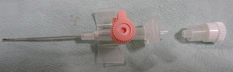
We attempted radial artery cannulation in left hand with 20G cannula (BD VenflonTM Pro IV Cannula, Becton Dickinson, Helsingborg, Sweden) in a patient with pancreatitis. First attempt was unsuccessful in negotiating the cannula into the artery. With repeated attempts radial artery went into spasm and pulse disappeared. Before trying to another artery we applied a simple modification to the technique in the same artery. We punctured the artery with a new cannula after distal white stopper removal (Figure 1) at the previous puncture point.
Figure1: Cannula with separated distal white stopper
As blood came into the hub, we tried to negotiate the catheter as much as possible till free arterial flow was there. Then we attached 2cc syringe filled with preservative free 2% lignocaine to distal hub of the cannula with or without stylet (Figure 2) and injected 1-1.5ml into artery and left the cannula for 45-60 sec.
Figure 2: Preservative free lignocaine filled 2 ml syringe at the distal end injecting as lignocaine stylet
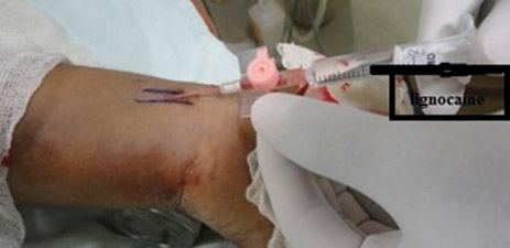
Figure 3: Cannula with stylet without stopper. Small drops of blood trickling without pulsatile flow
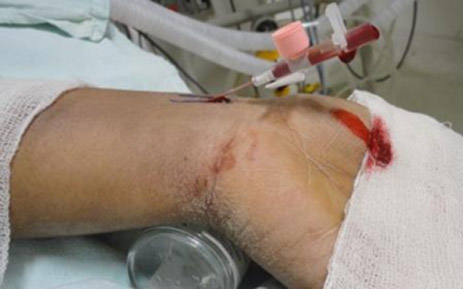
We could able to thread the cannula into the artery after the stipulated time successfully. Some people remove stylet to check arterial flow after puncturing artery with cannula. It causes profuse bleeding and soiling of the bed as sometimes it is difficult to reintroduce the stylet into cannula. To overcome this problem our suggestion is to remove the white stopper from the distal end of cannula and withdraw the stylet only a few milimeter before reducing angle to check free blood flow and threading while keeping stylet inside cannula. If the cannula with stylet is inside the artery, continuous small drops of blood will trickle from the stylet opening due to arterial pressure effect (Figure 3) but no trickling if the cannula is inside the vein. Thus it reduces blood loss with minimum soiling. We describe our modification as ‘Lignocaine stylet’ technique.
A “liquid-stylet” created by a slow arterial injection of saline through the catheter from distal end attached with saline filled syringe, has also been shown to be effective for insertion of arterial cannula.2 Different methods of identification of arteries like, hypodermic needle localization,3 and last ultrasound-guided arterial puncture have also been reported.4 But none will be effective when artery is in spasm. We have applied ‘Lignocaine stylet’ method in different arteries during difficult cannulation and got favourable results. It has been reported that vasospasm of carotid artery during aneurysm embolization surgery can be relieved by lidocaine infusion.5 Lignocaine can cause vasodilatation by blocking sodium channel in sympathetic nerves as well as release of nitric oxide from endothelium.6This method may also be helpful in children, infants and neonates with the use of 24G or 26G cannula and small volume of lignocaine 1% solution to limit the toxic dose. We have tried with other different agents like papavarine, nimodipine but lignocaine is promising and easily available with rapid onset of action.
So, we suggest anesthesiologists and intensivists to try this technique in spasmodic artery with difficult cannulation.
References
- Tegtmeyer K, Brady G, Lai S, Hodo R, Braner D. Videos in Clinical Medicine. Placement of an arterial line. N Engl J Med 2006;13:354. [PubMed][Free Full Text]
- Stirt JA. Liquid stylet for percutaneous radial artery cannulation. Can Anaes Svc J 1982;29:492-3. [PubMed]
- Penland WA, Henthorn RW. A method to assist radial artery cannulation. AnesthAnalg 1993;77:643. [PubMed]
- Schwemmer U, Arzet HA, Trautner H, Rauch S, Roewer N, Greim CA. Ultrasound guided arterial cannulation in infants improves success rate. Eur J Anaesthesiol 2006;23:476-80. [PubMed]
- Li YQ, Xu LW, Zhang Y, Lu PS, Yuan ZC, Zhan LP, et al. Intravascular Infusion of Lidocaine: A Novel Way to Relieve Sudden Internal Carotid Artery Occlusion in Embolization of Intracranial Aneurysms. J Neurol Surg A Cent Eur Neurosurg. 2012;73:84-8. [PubMed]
- Newton D McLeod GA, Khan F, Belch JJ.Mechanisms influencing the vasoactive effects of lidocaine in human skin. Anaesthesia. 2007;62:146-50. [PubMed]
