
Sacral bulge after double epidural space localization efforts in pediatric patients
To the editor,
Various techniques have been described for identification of the epidural space.1 We describe an interesting finding of sacral bulge during identifying epidural space in pediatric patients.
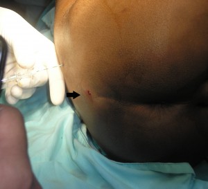
A 5 year old, ASA II, 15kgs, child was posted for elective posterolateral thoracotomy for empyema thoracis. Child was induced with intravenous fentanyl and propofol, and tracheal intubation was achieved using atracurium as muscle relaxant. To provide postoperative analgesia, an epidural infusion of low dose bupivacaine was planned. We decided to pass 24 G epidural catheter through caudal approach. Under aseptic conditions, with patient in lateral position, sacral epidural space was identified using 20 G Tuohy’s needle. We failed to thread the catheter even after two attempts We decided to pass a lumbar catheter instead. Same needle was introduced in L4-5 space using loss of resistance to air technique. When the air was pushed in, we could see an obvious and evident bulge with air leak in the sacral hiatus region (Figure-1) probably due to air escaping through the hiatal opening in the subcutaneous tissue. Catheter was secured and a compression dressing was applied. The surgery was uneventful and post operatively epidural infusion of low dose bupivacaine with opioids was given for three days and then the catheter was removed. The child was followed up and discharged on 15th post operative day.
Several techniques as well as different types of devices (viz, Page’s giving way method, Dogliotti’s loss of resistance technique, Gutierrez’s hanging drop method, Baraka’s running infusion, Cork’s ultrasonic method, Odom’s indicator, McIntosh Balloon, Brunner’s spring loaded plunger, Sagarnaga’s bursting bubbles) have been described over the years for identifying the epidural space.1 Most of these methods are based on the principle of demonstration of sub atmospheric pressure or sudden loss of resistance. In children, small anatomical structures and catheter insertion under general anesthesia poses difficulty to identify epidural space.2 We believe this ‘Caudal Bulge sign’ observed by us, though accidently, is more evident in thin patients.
Various complications associated with the use of air for the loss of resistance technique are pneumocephalus, spinal cord and nerve root compression, retroperitoneal air, subcutaneous emphysema, venous air embolism and inadequate analgesia and paresthesia.3 Air is no longer used for LOR in infants and children due to these risks. The technique described by us utilizes two puncture sites and may have potential to decrease the amount of air retained in the space, hence probably reducing the air related complications. Perhaps our observation of ‘sacral bulge sign’ may increase the reliability of loss of resistance technique in pediatric patients, though it requires double puncture. However, we on no account recommend using air in LOR technique or making two punctures to identify the space, but believe that the observation may make an interesting subject for further studies.
Figure 1: Figure showing sacral bulge after loss of resistance
REFERENCES
- Jacob S, Tierney E. A dual technique foe identification of the epidural space. Anaesthesia 1997;52:141-3
- Rapp HJ, Folger A, Grau T. Ultrasound-guided epidural catheter insertion in children. Anesth Analg. 2005;101:333-9
- Saberski LR, Kondamuri S, Osinubi OY. Identification of the epidural space: is loss of resistance to air a safe technique? A review of the complications related to the use of air. Reg Anesth. 1997;22:3-15
Dr. Uday Ambi, Assistant Professor, Dept. of Anaesthesiology, S N Medical College, Bagalkot, Karnataka-587102 (India); Phone- +91 9880570780; E-mail: udayambi@ymail.com
Misconnected epidural infusion into central line: A perfect recipe for disaster
Dear Editor,
Based on the severity of the sickness, patients may have several tubes and lines connecting them to devices for delivery of various drugs or nutrition through different routes. This increases the chance of misconnections in tubes and catheters, and may be potentially fatal. A major cause for these erroneous connections is the incorporation of the Luer lock, which permits the linking of functionally dissimilar tubes. Other causes include the use of tubes or catheters for unintended purposes (intravenous extension tubing for epidurals, irrigation, drains, and central lines, or to extend enteral feeding tubes), positioning of functionally dissimilar tubes in close proximity to one another, and movement of the patient from one setting or service to another.1 We came across such a potentially dangerous situation, where the extension tubing of the syringe containing epidural drug was inadvertently connected with one of the lumens of a central line.
A 62-year-old patient underwent Whipple’s procedure under combined general and epidural anaesthesia. After shifting the patient to the post anesthesia care unit, various lines and tubings were attached by the on-duty nurse. At the time of hand-over, it was discovered that the extension tubing for connecting the syringe containing the epidural solution (50 ml solution of 0.25% ropivacaine and 2 mcg/ml fentanyl), had been attached with one of the ports of the double lumen central line via another smaller extension tubing. This smaller extension tubing (20 cm) was attached to the central line for CVP monitoring. Through this connection, the epidural solution had been running intravenously for the past three hours at the rate of 5 ml/hr. The infusion was immediately stopped, and the extension tubing disconnected. The patient was monitored for any signs of toxicity and a close watch was kept on the vitals of the patient. Fortunately, no untoward incident occurred.
The Sentinel Event Alert, issued by Joint Commission in 2006, had cited different tubing and catheter misconnections, leading to eight deaths and one permanent loss of function.2 We were fortunate that no serious harm occurred to our patient, probably because the dose of ropivacaine (37.5 mg), administered inadvertently, was low. Moreover, the extension tubing, which was not primed with the epidural solution, would have accommodated some amount of the drug. Ropivacaine itself provided a degree of safety due to its lesser central nervous system and cardiovascular system effects in humans, as compared to bupivacaine.3 This incident emphasizes the necessity of anaesthesia providers to be extremely vigilant during connecting disconnected tubings, during and after transport. Rechecking and tracing of tubings to their origin, as a part of hand over process, saved the day for us. We would also like to voice our support for the recommendations of equipment design solution, e.g. labeling of high-risk catheter, routing tubings with standardized directions and limiting the use of adapters.4 The risk reduction strategy of labeling or color-coding feeding tubes and connectors, with necessary modifications, and educating the staff on this system,5 needs to be given due consideration.
References:
- The Joint Commission, Joint Commission International, World Health Organisation. Avoiding catheter and tubing mis-connections [Internet]. WHO Press;2007. Available from: http://www.who.int/patientsafety/solutions/patientsafety/PS-Solution7.pdf.
- The Joint Commission. Tubing misconnections—a persistent and potentially deadly occurrence [Internet]. Sentinel Event Alert;2006 Apr 3 [cited 2009 Nov 4]. Available from: http://www.jointcommission.org/assets/1/18/SEA_36.PDF
- Knudsen K, Beckman Suurküla M, Blomberg S, Sjövall J, Edvardsson N. Central nervous and cardiovascular effects of IV infusions of ropivacaine, bupivacaine and placebo in volunteers. Br J Anaesth. 1997;78(5):507-14. [Medline]
- Pennysylvania Patient Safety Advisory. Tubing misconnections: making the connection to patient safety [Internet]. Pa Patient Saf Advis 2010 Jun;7(2):41-5. Available from: http://patientsafetyauthority.org/ADVISORIES/AdvisoryLibrary/2010/Jun7(2)/Pages/41.aspx.
- Guenter P, Hicks RW, Simmons D, Crowley J, Joseph S, Croteau R et al. Enteral feeding misconnections: a consortium position statement. Jt Comm J Qual Patient Saf 2008 May;34(5):285-92,245.
Rudrashish Haldar, MBBS, MD, PDCC and Prakhar Gyanesh, MBBS, MD, PDCC
Dept of Anaesthesiology, SGPGI, Lucknow (India); rudrashish@yahoo.com
