
Şaban Cem Sezen1, Berrin Işık2, Mustafa Bilge3, Mustafa Arslan2, Faruk Metin Çomu4, Levent Öztürk5, Elvin Kesimci5, Mustafa Kavutçu3
Departments of 1Histology and Embryology and 4 Physiology, Faculty of Medicine, Kirikkale University, Kirikkale (Turkey)
Departments of 2Anesthesiology and Reanimation and 3Medical Biochemistry, Faculty of Medicine, Gazi University, Ankara (Turkey)
5Department of Anesthesiology and Reanimation, Ankara Atatürk Training and Research Hospital, Ankara (Turkey)
Correspondence: Dr. Mustafa Arslan, Gazi University Medical Faculty, Department of Anesthesiology and Reanimation, 06510 Ankara-Türkiye; Tel: 90 312 202 67 39; Fax: 90 312 202 4166; (GSM) 90 533 422 85 77; E-mail: marslan36@yahoo.com; mustarslan@gmail.com
ABSTRACT
Background: Reperfusion following ischemia can lead to more injuries than ischemia itself especially in diabetic patients. The aim of this study was to evaluate the effect of dexmedetomidine on ischemia-reperfusion injury (IRI) in rats with have hepatic IRI and diabetes mellitus.
Methodology: Twenty-eight Wistar Albino rats were randomised into four groups as control (C), diabetic (DC), diabetic with hepatic ischemia-reperfusion injury (DIR), and diabetic but administered dexmedetomidine followed by hepatic IRI (DIRD) groups. Hepatic tissue samples were evaluated histopathologically by semiquantitative methods. Malondialdehyde (MDA), superoxide dismutase (SOD), glutathion s-transpherase (GST), and catalase (CAT) enzyme levels were investigated in liver and kidney tissues as oxidative state parameters.
Results: In Group DIR; hepatocyte degeneration, sinusoidal dilatation, pycnotic nucleus, and necrotic cells were found to be more in rat hepatic tissue; while mononuclear cell infiltration was higher in the parenchyme. MDA levels were significantly lower; but SOD levels were significantly higher in Group DIRD with regard to Group DIR. In the IRI induced diabetic rats’ hepatic and nephrotic tissues MDA levels, showing oxidative injury, were found to be lower. SOD levels, showing early antioxidant activity, were higher.
Conclusion: The enzymatic findings of our study together with the hepatic histopathology indicate that dexmedetomidine has a potential role to decrease IRI.
Key words: Hepatic ischemia reperfusion injury; Diabetes mellitus; Dexmedetomidine; Rat; MDA; SOD
Citation: Sezen SC, Işık B, Bilge M, Arslan M, Çomu FM, Öztürk L, Kesimci E, Kavutçu M. Effect of dexmedetomidine on ischemia-reperfusion injury of liver and kidney tissues in experimental diabetes and hepatic ischemia-reperfusion injury induced rats. Anaesth Pain & Intensive Care 2016;20(2):143-149
Received: 21 November 2015; Reviewed: 10, 24 December 2015, 9, 10 June 2016; Corrected: 12 December 2015; Accepted: 10 June 2016
INTRODUCTİON
Perioperative acute tissue injury induced by ischemia-reperfusion is a comman clinical event caused by reduced blood supply to the tissue being compromised during major surgery. Ischemia leads to cellular injury by depleting cellular energy deposits and resulting in accumulation of toxic metabolites. The reperfusion of tissues that have remained in ischemic conditions causes even more damage.1 Furthermore hepatic ischemia-reperfusion injury (IRI) demonstrates a strong relationship with peri-operative acute kidney injury.2
The etiology of diabetic complications is strongly associated with increased oxidative stress (OS). Diabetic patients are known to have a high risk of developing OS or IRI which results with tissue failure.3
The most important role in ischemia and reperfusion is played by free oxygen radicals.1 In diabetes, characterized by hyperglycemia, even more free oxygen radicals are produced due to oxidation of glucose and glycosylation of proteins.3 The structures which are most sensitive to free oxygen radicals in the cells are membrane lipids, proteins, nucleic acids and deoxyribonucleic acids.1
It has been reported that endogenous antioxidant enzymes [superoxide dismutase (SOD), glutathion s-transpherase (GST), catalase (CAT)] play an important role to alleviate IRI.4-8 Also some pharmacological agents have certain effects on IRI.1 The anesthetic agents influence endogenous antioxidant systems and free oxygen radical formation.9-12
Dexmedetomidine is a selective α-2 adrenoceptor agonist agent. It has been described as a useful and safe adjunct in many clinical applications. It has been found that it may increase urine output by considerably redistributing cardiac output, inhibiting vasopressin secretion and maintaining renal blood flow and glomerular filtration. Previous studies demonstrated that dexmedetomidine provides protection against renal, focal cerebral, cardiac, testicular, and tourniquet-induced IRI.13-18 Arslan et al observed that dexmedetomidine protected against lipid peroxidation and cellular membrane alterations in hepatic IRI, when given before induction of ischemia.17 Si et al18 demonstrated that dexmedetomidine treatment results in a partial but significant attenuation of renal demage induced by IRI through the inactivation of JAK/STAT signaling pathway in an in vivo model.
The efficacy of the dexmedetomidine for IRI in diabetic patient is not resarched yet. The purpose of this experimental study was to evaluate the biochemical and histological effects of dexmedetomidine on hepatic IRI in diabetic rat’s hepatic and renal tissue.
METHODOLOGY
Animals and Experimental Protocol: This study was conducted in the Physiology Laboratory of Kirikkale University upon the consent of the Experimental Animals Ethics Committee of Kirikkale University. All of the procedures were performed according to the accepted standards of the Guide for the Care and Use of Laboratory Animals.
In the study, 28 male Wistar Albino rats, weighing between 250 and 300 g, raised under the same environmental conditions, were used. The rats were kept under 20-21 oC at cycles of 12-hour daylight and 12-hour darkness and had free access to food until 2 hours before the anesthesia procedure. The animals were randomly separated into four groups, each containing 7 rats.
Diabetes was induced by a single intraperitoneal injection of streptozotocin (Sigma Chemical, St. Louis, MO, USA) at a dose of 65 mg/kg body weight. The blood glucose levels were measured at 72 hrs following this injection. Rats were classified as diabetic if their fasting blood glucose (FBG) levels exceeded 250 mg/dl, and only animals with FBGs of > 250 mg/dl were included in the diabetic groups (diabetes only, diabetes plus ischemia-reperfusion and diabetes plus dexmedetomidine-ischemia-reperfusion). The rats were kept alive 4 weeks after streptozotocin injection to allow development of chronic diabetes before they were exposed to ischemia-reperfusion.(19) The rats were weighed before the study.
Rats were anesthetized with intraperitoneal ketamine 100 mg/kg. The chest and abdomen were shaved and each animal was fixed in a supine position on the operating table. The abdomen was cleaned with 1% polyvinyl iodine and when dry, the operating field was covered with a sterile drape and median laparotomy was performed. There were four experimental groups (Group C (sham-control; n = 7), (Group DC (diabetes-sham-control; n = 7), Group DIR (diabetes-ischemia-reperfusion; n = 7), and Group DIRD (diabetes-ischemia-reperfusion-dexmedetomidine; n = 7).
Sham operation was performed on the rats in Group C and Group DC. The sham operation consisted of mobilization of the hepatic pedicle only. The rats in this group were sacrificed 90 min after the procedure. Hepatic I/R injury was induced in Groups DIR and DIRD respectively with hepatic pedicle clamping using a vascular clamp as in the previous study of Arslan et al.(17) After an ischemic period of 45 min, the vascular clamp was removed. A reperfusion period was maintained for 45 min. In Group DIRD, dexmedetomidine hydrochloride 100 μg/kg, (Precedex 100 μg/2 ml, Abbott®, Abbott Laboratory, North Chicago, Illinois, USA) was administrated via intraperitoneal route 30 minutes before surgery.
All the rats were given ketamine 100 mg/kg intraperitoneally and intracardiac blood samples were obtained. Preserving the tissue integrity by avoiding trauma, liver and renal biopsy samples were obtained.
Biochemical Analysis: The liver and renal tissues were first washed with cold deionized water to discard blood contamination and then homogenized in a homogenizer. Measurements on cell contest require an initial preparation of the tissues. The preparation procedure may involve grinding of the tissue in a ground glass tissue blender using a rotor driven by a simple electric motor. The homogenizer as a tissue blender similar to the typical kitchen blender is used to emulsify and pulverize the tissue (Heidolph Instruments GMBH & CO KGDiax 900 Germany®) at 1000 U for about 3 min. After centrifugation at 10,000 g for about 60 min, the upper clear layer was taken.
MDA levels were determined using the method of Van Ye et al,(20) based on the reaction of MDA with thiobarbituric acid (TBA). In the TBA test reaction, MDA and TBA react in acid pH to form a pink pigment with an absorption maximum at 532 nm. Arbitrary values obtained were compared with a series of standard solutions (1,1,3,3-tetraethoxypropane). Results were expressed as nmol/mg.protein.
Part of the homogenate was extracted in ethanol/chloroform mixture (5/3 v/v) to discard the lipid fraction, which caused interferences in the activity measurements of T-SOD, CAT and GST activities. After centrifugation at 10.000 x g for 60 min, the upper clear layer was removed and used for the T-SOD, CAT, GST enzyme activity measurement by methods as described by Durak et al21, Aebi22 and Habig et al23 respectively. One unit of SOD activity was defined as the enzyme protein amount causing 50% inhibition in NBTH2 reduction rate and result were expressed in U/mg protein. The CAT activity method is based on the measurement of absorbance decrease due to H2O2 consumption at 240 nm. The GST activity method is based on the measurement of absorbance changes at 340 nm due to formation of GSH-CDNB complex.
Histological determinations: Semiquantitative evaluation technique used by Abdel-Wahhab et al(24) was applied for interpreting the structural changes investigated in hepatic tissues of control and research groups. According to this, (-) (negative point) represents no structural change, while (+) (one positive point) represents mild, (++) (two positive points) medium and (+++) (three positive points) represents severe structural changes.
Statistical analysis: The Statistical Package for the Social Sciences (SPSS, Chicago, IL, USA) 20.0 softwre was used for the statistical analysis. Variations in oxidative state parameters, and histopathological examination between study groups were assessed using the Kruskal-Wallis test. The Bonferroni-adjusted Mann-Whitney U-test was used after significant Kruskal-Wallis to determine which groups differed from the others. Results were expressed as mean ± standard deviation (Mean ± SD). Statistical significance was set at a p value < 0.05 for all analyses.
RESULTS
There was statistically significant difference observed between the groups with respect to findings from the histological changes in the rat liver tissue (hepatocyte degeneration, sinüsoidal dilatation, pycnotic nucleus, prenecrotic cell) determined by light microscopy according to semiquantitative evaluation techniques (p < 0.0001). In Group DIR, hepatocyte degeneration was significantly high compared to Group C, Group DC and Group DIRD (p < 0.0001, p < 0.0001, p = 0.002, respectively), (Table 1, Figure 1-4). Similarly, sinüsoidal dilatation was significantly higher in Group DIR (p < 0.0001, p = 0.004, p = 0.015, respectively). Although, pcynotic nucleus was decreased in Group DIRD, it did not make a significant difference in comparison to Group DIR (p = 0.053), (Table 1, Figure 1-4). The prenecrotic cells were significantly increased in Group DIR, with respect to Group C, Group DC and Group DIRD (p < 0.0001, p = 0.004, p < 0.0001, respectively), (Table 1, Figure 1-4).
Table 1. The comparison of histological changes in rat hepatic tissue [Mean ± SD)]
| Histological change | Group C
(n = 7) |
Group DC
(n = 7) |
Group DIR
(n = 7) |
Group DIRD
(n = 7) |
P** |
| Hepatocyte degeneration | 0.33 ± 0.52* | 0.50 ± 0.55* | 2.67 ± 0.57 | 1.17 ± 0.75* | < 0.0001 |
| Sinusoidal dilatation | 0.33 ± 0.55* | 1.00 ± 0.63* | 2.17 ± 0.41 | 1.17 ± 0.41* | < 0.0001 |
| Pycnotic nucleus | 0.0 ± 0.0* | 1.17 ± 0.41* | 2.17 ± 0.41 | 1.50 ± 0.55 | < 0.0001 |
| Cell pre necrosis | 0.0 ± 0.0* | 1.17 ± 0.41* | 1.83 ± 0.41 | 0.83 ± 0.41* | < 0.0001 |
| MN cellular infiltration in the parenchyma | 0.50 ± 0.55* | 1.33 ± 0.52* | 2.67 ± 0.82 | 1.33 ± 0.52* | < 0.0001 |
p**: Statistical significance was set at a p value < 0.05 for Kruskal-Wallis test
*p < 0.05: When compared with Group DIR
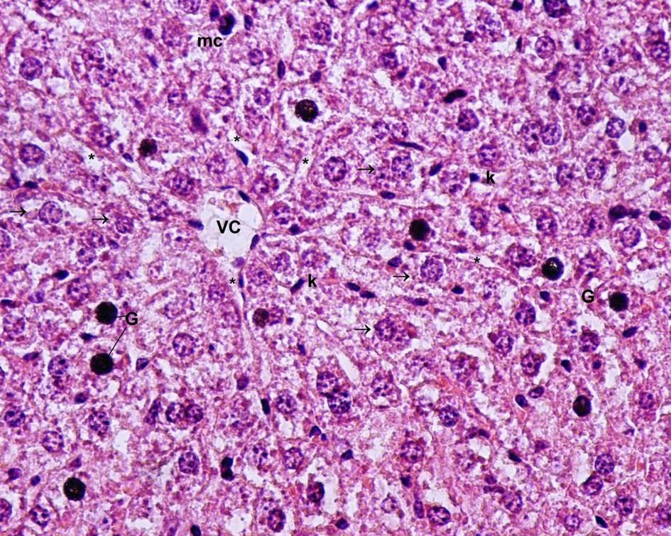
Figure 1: Light microscopic view of hepatic tissue of Group C (control). VC: vena centralis, *: sinusoids. ®: hepatocytes, k: Kupffer cells, G: glycogen granules, mc: minimal cellular changes, Hematoxilen & Eosin x 40
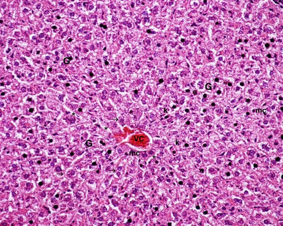
Figure 2: Light-microscopic view of hepatic tissue of Group DC (diabetes mellitus control) (G: Glycogen granules increased in number, (VC: vena centralis, *:sinusoids. ®:hepatocytes, k:Kupffer cells, G: glycogen granules, mc: minimal cellular changes; Hematoxylin & Eosin x 40)
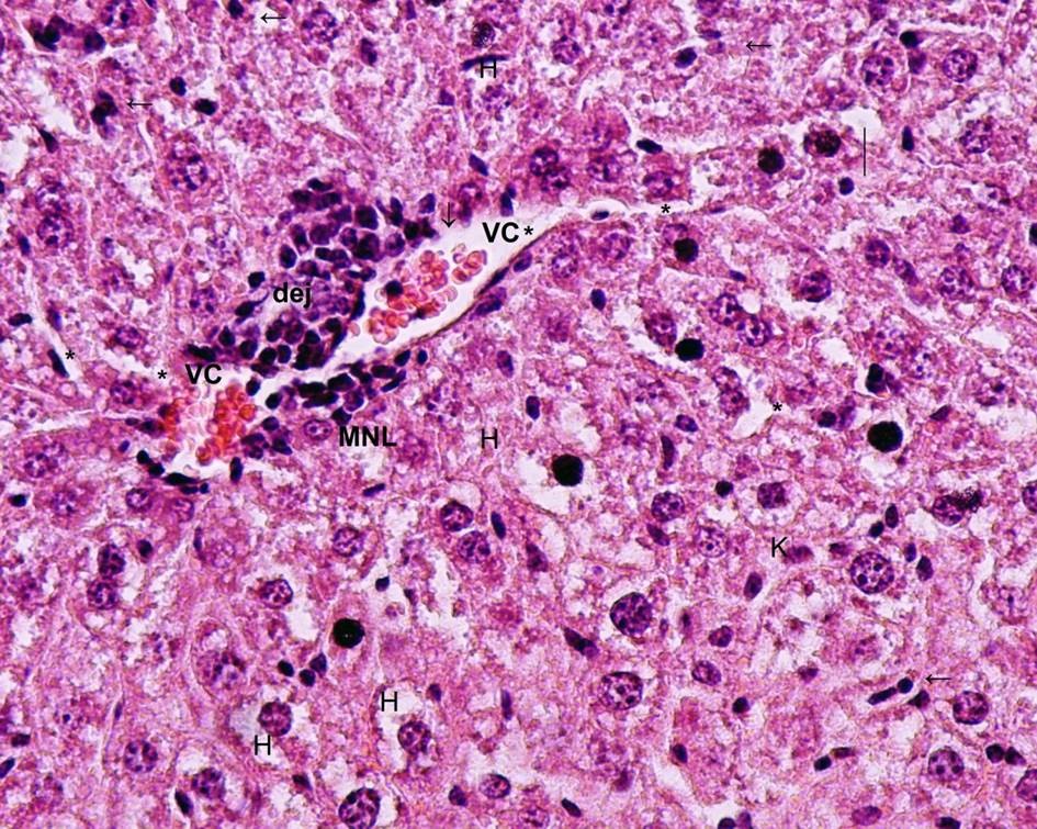
Figure 3: Light-microscopic view of hepatic tissue of Group DIR (Diabetes Mellitus and ischemia-reperfusion) (VC: vena centralis, (H) degenerative and hydrophic hepatocytes, (dej) vena centralis degeneration (centrolobar injury) (*): sinusoid dilatation. (←) pycnotic and hyperchromatic nuclei, MNL: mononuclear cell infiltration, (¯) congestion, K: Kupffer cell hyperplasia, () vacuolar degeneration (Hematoxylin & Eosin x 40)
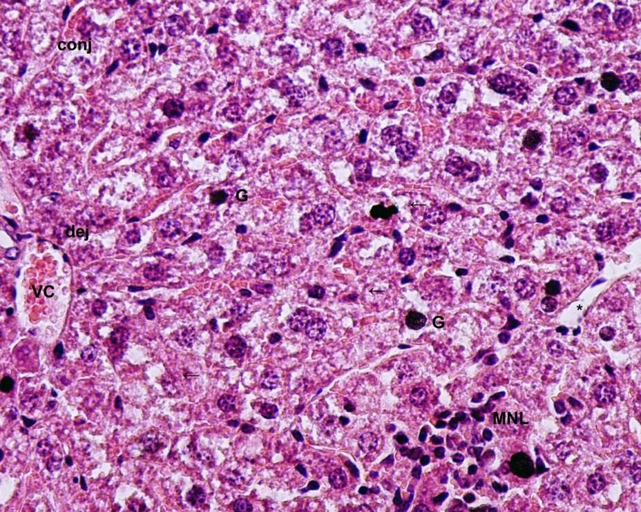
Figure 4: Light-microscopic view of hepatic tissue of Group DIRD (Diabetes Mellitus and ischemia-reperfusion together with dexmedetomidine applied group) (VC: vena centralis, (MNL) mononuclear cell infiltration, (dej) hydrophilic degeneration in hepatocytes around vena centralis, (conj) congestion, G: glycogen granules, (←) pycnotic and hyperchromatic nuclei, sinusoid dilatation (*) (Hematoxylin & Eosin x 40)
Besides, in liver tissue parenchyma, MN cellular infiltration was a light microscopic finding; and showed significant changes among the groups (p < 0.0001). This was significantly higher in Group DIR, compared to Group C, DC, and DIRD (p < 0.0001, p=0.007, p = 0.007, respectively), (Table 1, Figure 1-4).
The enzymatic activity of MDA, SOD and GST in hepatic tissues showed significant differences among the groups [(p = 0.019), (p = 0.034). (p = 0.008) respectively]. MDA enzyme activity was significantly incresed in Group DIR, according to Group C and Group DIRD (p = 0.011, p = 0.016, respectively), (Table 2). In Group DIR SOD enzyme activity was lower with respect to Group C and Group DIRD (p = 0.010, p = 0.038, respectively), (Table 2). The GST enzyme activity was significantly higher in Group DIR, when compared to Group C, DC and DIRD (p = 0.007, p = 0.038, p = 0.039, respectively), (Table 2).
Table 2. Oxidative state parameters in rat hepatic tissue [Mean ± SD]
| Parameters | Group C
(n = 7) |
Group DC
(n = 7) |
Group DIR
(n = 7) |
Group DIRD
(n = 7) |
P** |
| MDA (nmol/mg protein) | 0.23 ± 0.10* | 0.33 ± 0.11 | 0.55 ± 0.33 | 0.20 ± 0.07* | 0.019 |
| SOD (IU/mg protein) | 5.70 ± 1.36* | 3.02 ± 1.65 | 2.86 ± 1.55 | 5.42 ± 2.50 | 0.034 |
| GST (Liver) (IU/mg protein) | 0.97 ± 0.45* | 1.75 ± 0.66* | 3.58 ± 1.82 | 2.00 ± 0.91* | 0.008 |
| CAT (IU/mg protein) | 5038.39 ± 892.41 | 5614.72 ± 2057.04 | 6009.40 ± 857.38 | 5862.28 ± 940.41 | 0.289 |
p**: Statistical significance was set at a p value < 0.05 for Kruskal-Wallis test
*p < 0.05: When compared with Group DIR
The enzymatic activity of MDA, SOD in renal tissues, showed significant differences among the groups [(p < 0.0001), (p = 0.008) respectively ]. MDA enzyme activity was significantly incresed in Group DIR, according to Group C and Group DIRD (p < 0.0001, p < 0.0001, respectively). Also MDA enzyme activity level was significantly increased in Group DC, in comparison to Group C and Group DIRD (p = 0.003, p = 0.001, respectively), (Table 3). In Group DIR SOD enzyme activity was lower with respect to Group C and Group DIRD (p = 0.032, p = 0.013, respectively), (Table 3). The GST enzyme activity was significantly higher in Group DIR than the other three groups, however; CAT levels were similar among the groups (Table 3).
Table 3: Oxidative state parameters in rat nephrotic tissue [Mean ± SD)]
| Group C
(n = 7) |
Group DC
(n = 7) |
Group DIR
(n = 7) |
Group DIRD
(n = 7) |
P** | |
| MDA (nmol/mg prot) | 0.54 ± 0.08*.+ | 0.82 ± 0.07 | 0.86 ± 0.11 | 0.50 ± 0.14*.+ | < 0.0001 |
| SOD (IU/mg protein) | 3.20 ± 1.37* | 2.61 ± 0.57 | 1.27 ± 0.67 | 3.60 ± 1.25* | 0.008 |
| GST (Liver) (IU/mg prot) | 0.35 ± 0.08* | 0.40 ± 0.17* | 0.58 ± 0.12 | 0.40 ± 0.18* | 0.019 |
| CAT (IU/mg prot) | 2174.59 ± 1616.91 | 2885.16 ± 861.51 | 3210.84 ± 691.25 | 3047.49 ± 488.45 | 0.402 |
p**: Statistical significance was set at a p value < 0.05 for Kruskal-Wallis test
*p < 0.05: When compared with Group DIR
DISCUSSION
In this study, we have reported the protective effect of dexmedetomidine in experimental hepatic and renal IRI model in the rat by investigating the MDA and SOD levels biochemically. Besides, hepatic histopathological findings also supported our report. Ischemic damage may occur with trauma, hemorrhagic shock, and some surgical interventions, mainly hepatic and renal resections. Reperfusion following ischemia results in even more injury than ischemia itself.
IRI is an inflammatory response accompanied by free radical formation, leucocyte migration and activation, sinusoidal endothelial cellular damage, deteoriated microcirculation and coagulation and complement system activation.1 We also detected injury in hepatic and renal tissue caused by reperfusion following ischemia in liver.
Experimental and clinical evidence indicates that OS is involved in both the pathogenesis and the complications of diabetes mellitus.25,26 Diabetes mellitus is a serious risk factor for the development of renal and cardiovascular disease. It is also related to fatty changes in the liver.27 Diabetes-related organ damage seems to be the result of multiple mechanisms. Diabetes has been associated with increased free radical reactions and oxidant tissue damage in STZ-induced diabetic rats and also in patients.26 Oxidative stress has been implicated in the destruction of pancreatic β-cells28 and could largely contribute to the oxidant tissue damage associated with chronic hyperglycemia.29 A number of reports have shown that antioxidants can attenuate the complications of diabetes in patients30 and in experimental models.28,31 This study demonstrated that diabetes causes a tendency to increase the IRI.
There is a lot of investigations related to the pharmacological agents or food supplements applied for decreasing OS and IRI. Antioxidant agents paly an important role in IRI by effecting antioxidant system or lessening the formation of ROS. It has been reported that anesthetic agents too, are effective in oxidative stress.1 During surgical interventions, it seems rational to get benefit from anesthetic agents in prevention of OS caused by IRI instead of using other agents.
It has been declared that; dexmedetomidine; as an α-2 agonist with sedative, hypnotic properties; is important in prevention of renal, focal, cerebral, cardiac, testicular and tourniquet-induced IRI.13-18 On the other hand Bostankolu et al. concluded that dexmedetomidine did not have an additional protective role for tournique induced IRI during routine general anesthesia.32 In this study; we have shown that dexmedetomidine has a reducing effect in IRI in diabetic rats.
Some biochemical tests and histopathological evaluations are applied for bringing up oxidative stress and IRI in the tissues. Reactive oxygen species (ROS) that appear with reperfusion injury damage cellular structures through the process of the lipid peroxidation of cellular membranes and yield toxic metabolites such as MDA.33 As an important intermidiate product in lipid peroxidation, MDA is used as a sensitive marker of IRI.34 ROS-induced tissue injury is triggered by various defense mechanisms.35 The first defence mechanisms include the antioxidant enzymes of SOD, CAT, and GPx. These endogenous antioxidants are the first lines of defence against oxidative stres and act by scavenging potentially damaging free radical moieties.36 There is a balance between ROS and the scavenging capacity of antioxidant enzymes.1-8 In this study, for evaluation of oxidative damage and antioxidant activity, MDS, SOD, GST and CAT levels were determined in liver and kidney tissues. MDA levels in hepatic and renal tissues were higher in Group DIR compared to Group C and Group DIRD. GST levels were higher in Group DIR compared to all the other three groups. When the groups were arranged from highest to lowest order, with respect to CAT levels, the order was; Group DIR, Group DIRD, Group DC and Group C. However, the difference was not significant. The acute phase reactant MDA, as a marker of OS, was found to be high in Group DIR and low in Group DIRD. This could be interpreted as the presence of protective effect of dexmedetomidine in IRI.
IRI developing in splanchnic area causes injury also in the other organs.35 Leithead et al showed that clinically significant hepatic IRI demonstrates a strong relationship with peri-operative acute kidney injury.2 In our experimental research that showed correlation to that of research by Leithead et al. After hepatic IRI in diabetic rats renal OS marker MDA levels were significantly more in Group DIR than Group DIRD.
In our study, we observed histopathological changes in the ischemic liver tissue and alterations in the level of MDA, SOD, GST and CAT levels which are OS markers. Histopathological changes of the liver tissues are hepatocyt degeneration, sinusoidal dilatation, nuclear picnosis, celluler necrosis, mononuclear cell infiltrationat paranchimal tissue. These histopathological injury scores were significantly lower in the Group DIRD than those in group DIR.
LIMITATION
Study limitation is there was no negative control group, as this type of surgical intervention is not possible in rats without anesthesia.
CONCLUSION
The enzymatic findings of our study together with the hepatic histopathology indicate that dexmedetomidine has a potential role to decrease ischemia-reperfusion injury.
Conflict of interest and funding: The authors have not received any funding or benefits from industry or elsewhere to conduct this study.
Author contribution: ŞCS: Concept, conduction of the study work and manuscript editing; BI: the main author to write the article; MB & MK: biochemical analysis; MA: manuscript writing; FMÇ: helped us with experimental study; LÖ & EK: collection of data
REFERENCES
- Collard CD, Gelman S. Pathophysiology, clinical manifestations, and prevention of ischemia-reperfusion injury. Anesthesiology. 2001;94(6):1133. [PubMed] [Free full text]
- Leithead JA, Armstrong MJ, Corbett C, Andrew M, Kothari C, Gunson BK, et al. Hepatic ischemia reperfusion injury is associated with acute kidney injury following donation after brain death liver transplantation. Transpl Int. 2013;26(11):1116. doi: 10.1111/tri.12175. [PubMed] [Free full text]
- Panés J, Kurose I, Rodriguez-Vaca D, Anderson DC, Miyasaka M, Tso P, et al. Diabetes exacerbates inflammatory responses to ischemia-reperfusion. Circulation. 1996;93(1):161. [PubMed] [Free full text]
- Touyz RM. Reactive oxygen species and angiotensin II signaling in vascular cells-implications in cardiovascular disease. Braz J Med Biol Res. 2004;37:1263. [PubMed] [Free full text]
- Olivares-Corichi IM, Ceballos G, Ortega-Camarillo C, Guzman-Grenfell AM, Hicks JJ. Reactive oxygen species (ROS) induce chemical and structural changes on human insulin in vitro, including alterations in its immunoreactivity. Front Biosci. 2005;10:834. [PubMed]
- Witko-Sarsat V, Friedlander M, Capeillere-Blandin C, Nguyen-Khoa T, Nguyen AT, Zingraff J, et al. Advanced oxidation protein products as a novel marker of oxidative stress in uremia. Kidney Int. 1996;49:1304. [PubMed]
- Harman D. Free radical theory of aging: An update: Increasing the functional life span. Ann N Y Acad Sci. 2006;1067:10. [PubMed]
- Nita DA, Nita V, Spulber S, Moldovan M, Popa DP, Zagrean AM, Zagrean L. Oxidative damage following cerebral ischemia depends on reperfusion – a biochemical study in rat. J Cell Mol Med. 2001;5:163–170. [PubMed] [Free full text]
- Annecke T, Kubitz JC, Kahr S, Hilberath JM, Langer K, Kemming GI, et al. Effects of sevoflurane and propofol on ischaemia-reperfusion injury after thoracic-aortic occlusion in pigs. Br J Anaesth. 2007;98(5):581. [PubMed] [Free full text]
- De Hert SG, Van der Linden PJ, Cromheecke S, Meeus R, Nelis A, Van Reeth V, ten Broecke PW, et al. Cardioprotective properties of sevoflurane in patients undergoing coronary surgery with cardiopulmonary bypass are related to the modalities of its administration. Anesthesiology. 2004;101(2):299. [PubMed] [Free full text]
- Yuzer H, Yuzbasioglu MF, Ciralik H, Kurutas EB, Ozkan OV, Bulbuloglu E, et al. Effects of intravenous anesthetics on renal ischemia/reperfusion injury. Ren Fail. 2009;31(4):290. [PubMed] [Free full text]
- Lee HT, Ota-Setlik A, Fu Y, Nasr SH, Emala CW. Differential protective effects of volatile anesthetics against renal ischemia-reperfusion injury in vivo. Anesthesiology. 2004;101(6):1313. [PubMed] [Free full text]
- Lai YC, Tsai PS, Huang CJ. Effects of dexmedetomidine on regulating endotoxin-induced up-regulation of inflammatory molecules in murine macrophages. J Surg Res. 2009;154(2):212. doi: 10.1016/j.jss.2008.07.010. [PubMed]
- Yoshitomi O, Cho S, Hara T, Shibata I, Maekawa T, Ureshino H, Sumikawa K. Direct protective effects of dexmedetomidine against myocardial ischemia-reperfusion injury in anesthetized pigs. Shock. 2012;38(1):92. doi: 10.1097/SHK.0b013e318254d3fb. [PubMed]
- Jolkkonen J, Puurunen K, Koistinaho J, Kauppinen R, Haapalinna A, Nieminen L, et al. Neuroprotection by the alpha2-adrenoceptor agonist, dexmedetomidine, in rat focal cerebral ischemia. Eur J Pharmacol. 1999;372(1):31. [PubMed]
- Kocoglu H, Ozturk H, Ozturk H, Yilmaz F, Gulcu N. Effect of dexmedetomidine on ischemia-reperfusion injury in rat kidney: a histopathologic study. Ren Fail. 2009;31(1):70. doi: 10.1080/08860220802546487. [PubMed]
- Arslan M, Çomu FM, Küçük A, Öztürk L, Yaylak F. Dexmedetomidine protects against lipid peroxidation and erythrocyte deformability alterations in experimental hepatic ischemia reperfusion injury. Libyan J Med. 2012;7. doi: 10.3402/ljm.v7i0.18185 [PubMed] [Free full text]
- Si Y, Bao H, Han L, Shi H, Zhang Y, Xu L, et al. Dexmedetomidine protects against renal ischemia and reperfusion injury by inhibiting the JAK/STAT signaling activation. J Transl Med. 2013;11(1):141. doi: 10.1186/1479-5876-11-141. [PubMed] [Free full text]
- Türeci E, İş M, Üzüm G, Akyüz F, Ulu MO, Döşoğlu M, et al. Alterations in blood-brain barrier after traumatic brain injury in streptozotocin-induced diabetic rats. J Nervous Sys Surgery 2009;2(2):79. [Free full text]
- Van Ye TM, Roza AM, Pieper GM, Henderson J Jr, Johnson JP, Adams MB. Inhibition of intestinal lipid peroxidation does not minimize morphological damage. J Surg Res 1993;55:553. [PubMed]
- Durak I, Canbolat O, Kavutcu M, Öztürk HS, Yurtarslanı Z. Activities of total, cytoplasmic and mihochondrial superoxide dismutase enzymes in sera and pleural fluids from patient with lung cancer. J Clin Lab Anal 1996;10:17. [PubMed]
- Aebi H. Catalase. In: H.U.Bergmeyer (Ed): Methods of Enzymatic Analysis, Academic Press , New York and London, 1974;pp.673-677.
- Habig WH, Pabst MJ, Jakoby WB. Glutathione S-transferases. The first enzymatic step in mercapturic acid formation. J Biol Chem 1974;249:7130. [PubMed] [Free full text]
- Abdel-Wahhab MA, Nada SA, Arbid MS. Ochratoxicosis: Prevention of developmental toxicity by L-methionine in rats. J Appl Toxicol 1999;19:7. [PubMed]
- Wolff SP. Diabetes mellitus and free radicals: free radicals, transition metals and oxidative stress in the aetiology of diabetes mellitus and complications. Br Med Bull. 1993;49:642. [PubMed] [Free full text]
- West IC. Radicals and oxidative stress in diabetes. Diabet Med. 2000;17:171–180. [PubMed]
- Wanless IR, Lentz JS. Fatty liver hepatitis (steatohepatitis) and obesity: an autopsy study with analysis risk factors. Hepatology. 1990;12:1106. [PubMed]
- Hotta M, Tashiro F, Ikegami H, Niwa H, Ogihara T, Yodoi J, Miyazaki J. Pancreatic cell-specific expression of thioredoxin, an antioxidative and antiapoptotic protein, prevents autoimmune and streptozotocin-induced diabetes. J Exp Med. 1998;188:1445. [PubMed] [Free full text]
- Baynes JW. Role of oxidative stress in the development of complications in diabetes. Diabetes. 1991;40:405. [PubMed]
- Borcea V, Nourooz-Zadeh J, Wolff SP, Klevesath M, Hofmann M, Urich H, et al. α-Lipoic acid decreases oxidative stress even in diabetic patients with poor glycemic control and albuminuria. Free Radic Biol Med. 1999;26:1495. [PubMed]
- Fitzl G, Martin R, Dettmer D, Hermsdorf V, Drews H, Welt K. Protective effect of ginkgo biloba extract EGb 761 on myocardium of experimentally diabetic rats, I: ultrastructural and biochemical investigation on cardiomyocytes. Exp Toxicol Pathol. 1999;51:189. [PubMed]
- Bostankolu E, Ayoglu H, Yurtlu S, Okyay RD, Erdogan G, Deniz Y, et al. Dexmedetomidine did not reduce the effects of tourniquet-induced ischemia-reperfusion injury during general anesthesia. Kaohsiung J Med Sci. 2013;29(2):75. doi: 10.1016/j.kjms.2012.08.013. [PubMed] [Free full text]
- Wakai A, Wang JH, Winter DC, Street JT, O’Sullivan RG, Redmond HP. Tourniquet-induced systemic inflammatory response in extremity surgery. J Trauma 2001;51:922. [PubMed]
- Concannon MJ, Kester CG, Welsh CF, Puckett CL. Patterns of free-radical production after tourniquet ischemia implications for the hand surgeon. Plast Reconstr Surg 1992;89:846. [PubMed]
- Grisham MB, Granger DN. Free radicals: reactive metabolites of oxygen as mediators of postischemic reperfusion injury. In: Martson A, Bulkley GB, Fiddian-Green RG, Haglund U, editors. Splanchnic İschemia and Multiple Organ Failure. St Louis, MO: Mosby;1989. pp. 135–144.
- Mccard JM. The evolution of free radicals and oxidative stress. Am J Med 2000;108:652. [PubMed]
