
Nighat Abbas1, Arif Iftikhar Mallhi2, Syed Sajjad Alam3, H.M. Abid Mughal3
1Professor / HoD; 2Consultant Anesthetist; 3Resident Medical Officer
Department of Anesthesiology, Liaquat National Hospital and Medical College, Karachi (Pakistan)
Correspondence: Dr Syed Sajjad Alam, Resident Medical Officer Anaesthesia, Liaquat National Hospital and Medical College Karachi-Pakistan; E-mail: sajjadalamdr15@gmail.com; Phone: 0333-3607488
ABSTRACT
A 5 days old baby was referred from a local hospital for evaluation and treatment of a huge swelling on the right side of the neck extending beyond the midline in front and with difficulty by her mother to feed her since birth. It was clinically diagnosed to be cystic hygroma and a decision was made to further evaluate by doing magnetic resonance imaging under general anesthesia in order to see the extent of the mass and to plan for either sclerotherapy or surgical excision. Difficult intubation was anticipated and the baby was intubated in main operating room with inhalation induction and maintenance with sevoflurane without giving muscle relaxants. The 30 min of the procedure period was smooth without any complication. The baby was extubated in the main operating room complex.
Key words: Cystic hygroma; Difficult airway; Neonate
Citation: Abbas A, Mallhi AI, Alam SS, Mughal HMA. Cystic hygroma, difficult airway and the anesthetic considerations. Anaesth Pain & Intensive Care 2017;21(4):472-474
Received: 15 May 2017; Reviewed: 4 Jun, 22 Jun 2017; Corrected: 11 Aug 2017; Accepted : 15 Aug 2017
INTRODUCTION
Hygroma is a Greek word which means water containing tumor. Cystic hygroma is a benign tumor which is composed of large cysts and classified as capillary, cavernous and cystic lymphangiomas. It is also classified on the basis of size of the cysts as microcystic, macrocystic and mixed lymphangiomas. A microcystic lymphangioma consists of cysts < 2 cm, macrocystic lymphangioma consist of cysts > 2 cm and mixed lymphangiomas consist of cysts of variable sizes.1,2 The airway and anesthetic management in neonates and adults has many significant differences. Maintaining the airway of neonate and young infants with large neck mass like a huge cystic hygroma can be a challenge for any anesthetist, as airway obstruction, hypoventilation and hypoxemia and sudden collapse are very common risk factors due to tumor extension in the neck, airway and thorax. We describe the difficulties encountered in intubation and anesthetic management in our neonatal patient.
CASE REPORT
A 5 days old baby girl of 2.8 kg delivered on term with spontaneous vacuum delivery in a local hospital of Karachi Pakistan. She had late cry for about 5 min, maintaining saturation 97% with oxygen of 5 L, heart rate of 150 beats/min and was referred for evaluation and treatment of a huge swelling on the right side of the neck extending beyond the midline in front. She had difficulty in feeding mother’s milk since birth. The swelling in the neck of fetus had been diagnosed during intrauterine life of 8 m gestation through fetal scan. It was small in size when noticed at birth, which progressed to the present size (Figures 1 & 2)..
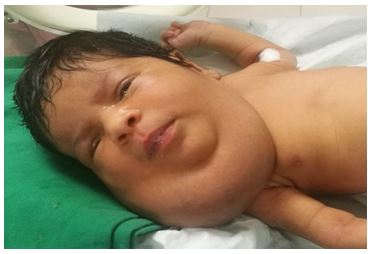
Figure 1: Anterior view of neck mass
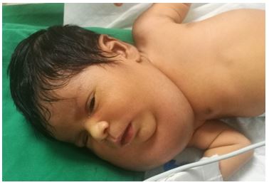
Figure 2: Anterolateral view of the mass
The child presented with a huge swelling with difficulty in feeding without restricted mouth opening. Examination revealed swelling on the right side of the neck measuring about 6.3×7.9×7.5 cm (AP×TS×LS), which was cystic, non-tender and extending from the angle of the right mandible to the right clavicle. The skin over the swelling looked normal with no local rise of temperature.
Pre-anesthesia assessment was carried out with systemic review i.e. electrocardiography was within normal limits and normal S1+S2+0 cardiac sounds on auscultation, chest was clear with normal vesicular breathing and equal air entry bilaterally on auscultation, thyromental distance was less than 3 fingers, labs (complete blood count, erythrocyte sedimentation rate etc.) were normal in range and chest x-ray was done to exclude the presence of chest infection and intrathoracic extension of the tumor.
Keeping in mind the suspected difficult airway we kept ready a difficult airway cart that contained face mask of various sizes, oropharyngeal airways of assorted sizes, nasopharyngeal airways, endotracheal tubes size 2.5, 3 and 3.5, stylet, suction, bougie, short handle laryngoscope, straight blade, videolaryngoscope, laryngeal mask airway No. 1 and 1.5.
The child was premedicated with inj glycopyrrolate 0.005 mg/kg (0.014 mg). A shoulder roll was used to keep the child at optimal laryngoscopic position and preoxygenated with high flow 100% oxygen into the breathing system in order to increase oxygen reserve which might allow more time for laryngoscopy and tracheal intubation.
The child was induced with sevoflurane in oxygen by an effective face mask seal. A straight blade laryngoscope was used as child had an anterior and cephalad placed larynx and epiglottis was difficult to be visualized. At first attempt, laryngoscopy was difficult as the epiglottis and vocal cords were not visualized due to displacement of soft tissue to the left side. The child was ventilated with mask and a second attempt at laryngoscopy was made by shifting the soft tissues to the right side. It helped to visualize the glottis with assistance and the trachea was successfully intubated with uncuffed endotracheal tube (ETT) size 3 mm; fixed just 1 cm beyond the vocal cords in order to avoid accidental extubation. The child was deeply sedated with sevoflurane showing no cough reflex and did not require any muscle relaxant.
The child was transported for magnetic resonance imaging (MRI) procedure room after taking transport consent with parents of the child in an incubator with monitoring all vitals and electrocardiography with assisted ventilation. After taking the child on MRI table, all vital signs were monitored with MRI compatible monitoring system and anesthesia was maintained with isoflurane in oxygen with controlled ventilation with Jackson Rees modification of Ayre’s t piece. At the end of procedure, the child was shifted back to the main operating room for extubation with all precautions taken during transportation to MRI room. The child was extubated after return of adequate muscle power, respiratory effort, good cry and limb movements.
DISCUSSION
Cystic hygroma, also called cavernous hemangioma, is caused by blockages in the lymphatic system and is a histological benign congenital tumor developed in 9th and 16th week of pregnancy.3,4
Embryologically, surrounding normal tissues are penetrated by sprouting endothelial membrane sequestered lymph vessels that forms fimbrillae causing canalization and production of large multiloculated cysts filled with serous secretion.5 The condition may develop due to some genetic disorder like Turner syndrome, trisomy 13,18 or 21, Noonan syndrome or may be due to environmental factors like viral infections, exposure to drugs or alcohol passed from mother to baby during pregnancy. Approximately half of all fetuses with a cystic hygroma have chromosomal abnormalities therefore other related abnormalities should also be ruled out.4
Cystic hygroma usually presents in neonates and during early infancy but can develop at birth or with obstructed labor.6 The most prominent sign of cystic hygroma is presence of a soft and spongy lump which commonly appears on neck that can interfere with normal breathing and swallowing.7
Securing the airway was the prime consideration in our case. A difficult airway is a clinical situation in which a conventionally trained anesthesiologist experiences difficulty with facemask ventilation, or difficulty with tracheal intubation or both. A difficult laryngoscopy is when it is impossible to view any part of the vocal cords during direct laryngoscopy. Managing a difficult airway in newborn can be a nightmare for any physician. Intubation and managing the secure airway in newborn is difficult, and the success ratio of intubation is only 60% on first attempt. In addition, lower functional residual capacity makes them prone to rapid development of hypoxia, so it is recommended to limit the duration of laryngoscopy and intubation to less than 20 sec.8 In babies with anticipated difficult airway intubation should be carried out under general anesthesia or deep sedation, preferably maintaining spontaneous ventilation until trachea is successfully intubated.9 Extubation should be done after return of adequate muscle power, respiratory efforts, response to commands, and when there is no cardiopulmonary instability, and the cry in case of newborn or infants is good.10
CONCLUSION
Difficult airway due to head and neck mass effect can be a challenge to any anesthetist. Inhalational induction is the preferable techniques. Endobronchial intubation along with intra-operative management of intubation, accidental extubation and anticipation of possible post-operative complications should be considered. A definite plan to deal with ‘can’t intubate, can’t ventilate’ (CICV) situations, availability of appropriate equipment and critical skills are fundamental for the airway management in such cases. Similarly, during extubation, intact airway reflexes, GCS level, the respiratory effort and postoperative management should be considered to improve the prognosis of the patient.
Conflict of interest: Nil
Authors’ Contribution:
NA – Supervised the case, manuscript editing & reviewing
AIM – pre-anesthesia assessment, conduction of anesthesia
SSA – Concept, assisted the case, manuscript writing, editing
HMAM – Assisted the case
REFERENCES
- Lymphatic disorders. In: Grosfeldjl, O’neilljajr, Coranja, Fonkalsrudew, Caldamoneaa, Editors. Pediatric Surgery. 6th ed. Chicago: Mosby Elsevier;2006. Pp. 2137–45.
- Sanlialp I, Karnak I, Tanyel FC, Senocak ME, Buyukpamukcu N. Sclerotherapy for lymphangioma in children. Int J Pediatrotorhynolaryngol. 2003;67:795–800. [PubMed]
- Das S. Textbook of Surgery. In: Das S, editor. 6th ed. Kolkata: SD Publishers; 2010. P. 507.
- April kahn. Your baby and cystic hygromas. Health line newsletter. 2015 December 1.
- Ward PH, Harris PF, Downey W. Surgical approach to cystic hygroma of the neck. Arch otolaryngol. 1970;91:508–14. [PubMed]
- Williams NS, Christopher JK, Ronan O’connell. 25th ed. London: Edward Arnold (publishers) ltd; 2008. Pharynx, larynx and neck.Bailey and love’s short practice of surgery; p. 279.
- Sharma S, Aminuldin AG, Azlan W. Cystic hygroma: anesthetic considerations and review. Singapore med j. 1994;35:529–31. [PubMed]
- Kim H, Kim HS, Oh JT, Lee JR. Anesthetic management for neonate with giant cystic hygroma involved upper airway:a case report. Korean j anesthesiol. 2011;60:209–13.[PubMed] [Free full text]
- Bryan Y, Chwals W, Ovassapian A. Sedation and fiberoptic intubation of a neonate with a cystic hygroma. Acta anaesthesiol scand. 2005;49:122–3. [PubMed]
- Whitten C. To Extubate, Or Not to Extubate, That Is The Question. Anyone Can Intubate. The Airway Jedi. [Online]. 2016 Jan 7. Available from: URL:https://airwayjedi.com/2016/01/07/to-extubate-or-not-to-extubate-that-is-the-question/https://airwayjedi.com/2016/01/07/to-extubate-or-not-to-extubate-that-is-the-question/
