
Arun Kumar Gupta*, V.K.Dhulkhed*, B.M.Rudagi**, Ana Gupta*
*Dept of Anesthesiology
** Dept of Oral & Maxillofacial Surgery
Rural Medical College, Loni, Maharashtra, (India). 413736
Correspondence: Dr.Arun Kumar Gupta, Assistant Professor, Dept of Anesthesiology, Rural Medical College, Loni, Maharashtra (India). 413736;
Phone no: +919823262700; Email: guptaarun71@gmail.com
ABSTRACT
Ludwig’s angina is rapidly spreading cellulitis that may result in potentially lethal upper airway obstruction. There is very little published literature regarding this condition in the pregnant patient. We present a case report of a 20 years old female patient admitted at 30 weeks gestation with tooth pain, submandibular swelling, severe trismus and dysphagia, consistent with Ludwig’s angina. Her planned treatment included incision and drainage of associated spaces, teeth extraction, and antibiotic therapy. During a life threatening infectious condition such as the one we describe, risks to the mother and the baby include septicemia, hypoxia and/or asphyxia. We successfully relieved airway obstruction by surgical decompression alone, using a cervical plexus block.
Keywords: Pregnancy; Ludwig’s angina; Cervical plexus block.
Citation: Gupta AK, Dhulkhed VK, Rudagi BM, Gupta A. Drainage of Ludwig’s angina in a pregnant patient under superficial cervical plexus block. Anaesth Pain & Intensive Care 2009;13(2):68-70
INTRODUCTION
Ludwig’s angina is defined as a potentially lethal, rapidly spreading cellulitis, involving the sublingual and submandibular spaces, and is manifested by a brawny suprahyoid induration, tender swelling in the floor of the mouth, and elevation and posterior displacement of the tongue.1
The most common cause of Ludwig’s angina is an odontogenic infection from one or more grossly decayed, infected teeth, and is usually as a result of native oral streptococci or a mixed aerobic-anaerobic oral flora.2 Prompt airway management is critical, but the presence of swelling of the neck, glottic edema, elevation of the tongue, trismus, or pharyngeal edema create formidable problems.3
It is a life threatening condition, yet an extensive literature search did not yield much published information regarding its occurrence in the pregnant patients.
CASE REPORT
A 20-year old primigravida, at 30 weeks gestation, presented to our hospital with complaints of facial swelling. She described that her pregnancy had been uneventful, except for a seven-day history of lower left quadrant tooth pain, and a three-day history of fever and chills. Her clinical examination revealed a large soft tissue swelling under her mandible, extending bilaterally to the angles of the mandible and inferiorly up to her hyoid bone. On presentation, her vital signs were: temperature 38.7°C, blood pressure 125/54, pulse 116/min, respiratory rate 19/min, oxygen saturation on room air 96%, and white cell count of 16800/μL. The diagnosis of Ludwig’s angina was made.
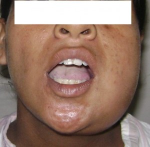
It was difficult to perform an adequate oral exam due to pain, swelling, and severe trismus which allowed her to open her mouth to only 15 mm (average range 40–45 mm). The patient had difficulty in containing her own salivary secretions because of dysphagia, but had no dyspnea. Since in similar situations patients may desaturate very quickly, even though her oxygen saturation was recorded to be 96% on room air, she was given supplemental oxygen and a pulse oximeter was attached. An emergent cricothyrotomy kit was kept available at the patient’s bedside at all times. We formally consulted the Department of Obstetrics and Gynecology, understanding that in administering medications and/or undergoing any surgical treatment in pregnancy, one must consider the risks and the benefits both to the mother and the unborn, it was determined that the benefits of proceeding with emergent and immediate surgical intervention outweighed the risks.
Figure 1: Limited mouth opening
Securing an airway via an awake fiberoptic nasal intubation was risky: a fiberoptic tube inserted into the pharynx might puncture an abscess and cause pus aspiration or swallowing. It was thus decided to attempt a trial of decompression under superficial cervical plexus block. Complete preparations for an emergency tracheostomy were also undertaken.
Standard monitors were attached. The patient was placed in a supine position, with her head turned to the right side. Under aseptic technique, lidocaine 1% was infiltrated at the midpoint of the line connecting the mastoid process with Chassaignac’s tubercle of C6 transverse process. Then inj.buvipacaine 0.5% 8ml was injected after negative aspiration using a fan technique along the posterior border of sternocleidomastoid muscle which reduced the pain and enabled the patient to open mouth more widely. An inferior alveolar nerve block was then performed by maxillofacial surgeon intraorally.
Dense anesthesia was established in about 7 min. A rapid decompression of the left submandibular region was done and the mylohyoid transected with resultant lowering of the floor of mouth, the blunt dissection continued through the mylohyoid muscle to the sublingual areas to access all loculations. Lower left three molars were then extracted since it was believed that these grossly carious and partially impacted teeth were the primary source of the infection. Upon removal, purulence was expressed through the extraction socket. There was little discharge from the wound, which was lightly packed and dressed. Post op fetal heart sound was monitored by fetal Doppler. After four days, she was discharged, to be followed up at Departments of Oral & Maxillofacial Surgery and Obstetrics & Gynecology.
DISCUSSION
The unique anatomy of the floor of the mouth plays an important role in the development and extension of intraoral infections. The usual infectious course begins with a periapical dental abscess of the second or third mandibular molar. The roots of these teeth extend inferior to the insertion of the mylohyoid muscle, so that if untreated, the infection may continue from primary spaces to penetrate the thin inner cortex of the mandible and will involve the posterior margin of the mylohyoid muscle to the submandibular space. At this point, the infection may develop and progress at such an alarming rate that special precautions regarding airway maintenance must be taken. 4
It is estimated that about 50,000 women require anesthesia and a surgical intervention each year at some time during gestation for indications unrelated to the pregnancy.5 In such situations, when medical and surgical treatments for pregnant women are considered, both the physiologic changes of pregnancy and the perinatal effects of the treatment must be considered.6 Pregnancy is accompanied by physiological changes which place the mother at a higher risk of infection or of doing worse once infected. First, the immune response is greatly diminished during pregnancy, thus resulting in rapid progression of an infection. Secondly, there is decreased neutrophil chemotaxis, cell mediated immunity, and natural killer cell activity.7,8 Moreover, approximately 50% of pregnant women complain of some degree of dyspnea by 19th week of gestation7 and there is some depletion in the oxygen reserve of the gravid patient. This could increase fetal hypoxia during periods of hypoventilation.6 From an oral perspective, as pregnancy associated hormonal changes begin to affect a woman’s body, the gingival tissues are affected as well. They become much more sensitive and thus susceptible to irritation from soft plaque. The plaque accumulates, becomes hard calculus deposits on the teeth, and harbors bacteria in large numbers resulting in a constant, low-grade intraoral infection. Maternal infective processes sustained especially by gram negative anaerobic bacteria, such as those leading to Ludwig’s angina, have been demonstrated to cause physiologic imbalance through inflammatory cytokine production, sometimes resulting in preterm labor, premature rupture of membranes, and low birth weight.9 During pregnancy, women tend to have frequent meals and snacks, which augment plaque accumulation, as well as an increase in decay or rapid progression of previously present decay. A remote infection can at times infect the placenta, uterus, and possibly the fetus, causing fetal septicemia. During a life threatening infectious situation such as the one described, the risk of maternal and fetal morbidity may overshadow potential teratogenic side effects during early pregnancy.10
In an exhaustive review of the literature, from 1945 to 1979, 75 cases of Ludwig’s angina were found, and the authors strongly advocate elective tracheostomy under local anaesthesia.11 Cellulitis of the neck with involvement of the tracheostomy site may make it a more difficult option. Moreover, surgical dissection of the fascial planes in the neck may actually open and contaminate the pathways, leading to life-threatening mediastinal invasion.12
Other options for airway management include orotracheal, blind nasotracheal, and fiber optic intubation or cricothyroidotomy with jet insufflation. We chose to employ a cervical plexus block as anesthesia for surgical decompression. The block permitted a thorough incision and drainage, including transection of mylohyoid with lowering of the floor of mouth and rapid relief of respiratory obstruction. Ling et al also recommended the consideration of superficial cervical plexus block, and if necessary an auriculotemporal nerve block to manage selected patients with difficult airways who present for drainage of dental abcesses.13 Moshe et al advocated superficial cervical plexus block with concomitant mandibular nerve block with a high success rate, low complication rate and high patient acceptance rate for the drainage of submandibular and submental abscesses.14
Ludwig’s angina is life threatening because of risks of septicemia and asphyxia. Furthermore, in pregnancy, the condition itself as well as possible therapies may put the mother and her unborn child at increased risk.
CONCLUSION
Superficial cervical plexus block combined with mandibular nerve block can safely be employed for the surgical decompression in a case of pregnant patient with Ludwig’s angina.
REFERENCES
- Patterson H, Kelly JH, Strone M: Ludwig’s angina: An update. Laryngoscope 1982;92:370.
- Topazian RG, Goldberg MH, Hupp JR. Oral and Maxillofacial Infections. 4th ed. Philadelphia, Pa: W. B. Saunders; 2002.
- Allen D, Loughnan TE, Ord RA: A reevaluation of the role of tracheostomy in Ludwig’s angina. J Oral Maxillofac Surg 1985;43:436-9.
- Marple BF. Ludwig angina: a review of current airway management. Archives of Otolaryngology – Head and Neck Surgery. 1999;125(5):596–600.
- Aroesty JH, Lanza JT, Lucente FE. Otolaryngology and pregnancy—difficult management decisions. Otolaryngology – Head and Neck Surgery. 1993;109(6):1061–1069.
- Barron WM. Medical evaluation of the pregnant patient requiring nonobstetric surgery. Clinics in Perinatology. 1985;12(3):481–496.
- Silver RM, Peltier MR, Branch DW. The immunology of pregnancy. In: Creasy RK, Resnik R, editors. Maternal-Fetal Medicine: Principles and Practice. Philadelphia, Pa: W. B. Saunders; 2004. pp. 89–109.
- Lawrenz DR, Whitley BD, Helfrick JF. Considerations in the management of maxillofacial infections in the pregnant patient. J Oral Maxillofac Surg. 1996;54(4):474–485.
- Scannapieco FA, Bush RB, Paju S. Periodontal disease as a risk factor for adverse pregnancy outcomes. A systematic review. Annals of Periodontology. 2003;8:70–78.
- Moore PA. Selecting drugs for the pregnant dental patient. Journal of the American Dental Association. 1998;129(9):1281–1286.
- Hought RT, Fitzgerald BE, Latta JE, Zallen RD: Ludwig’s angina: A report of two cases and review of the literature from 1945 to January 1979. J Oral Surg 1980;38:849-55.
- Snow N, Lucas AE, Grau M, Steiner M: Purulent mediastinal abscess secondary to Ludwig’s angina. Arch Otolaryngol 1983;109: 53-5.
- Ling KU, Hasan MS, Ha KO, Wang CY. Superficial cervical plexus block combined with auriculotemporal nerve block for drainage of dental abscess in adults with difficult airways. Anaesth Intensive Care 2009;37(1):124-6.
- Shteif M, Lesmes D, Hartman G, Ruffino S, Laster Z. The use of the superficial cervical plexus block in the drainage of submandibular and submental abscesses–an alternative for general anesthesia. J Oral Maxillofac Surg. 2008 Dec;66(12):2642-5.
