
Saurabh Kumar Das,MD*, D. K. Singh,MD**, Sujali Choupoo,MD*, Ghanshyam Yadav,MD***
*Senior Registrar; **Professor; ***Assistant Professor
Institute of Medical Science, Banaras Hindu University, Varanasi, Uttar Pradesh (India)
Correspondence: Dr Saurabh Kumar Das, Intensive Care Unit, SS Hospital, Institute of Medical Sciences, Banaras Hindu University, Varanasi, Uttar Pradesh (India); E-mail – drsauravdas1977@gmail.com
ABSTRACT
Obesity, sleep apnea syndrome and hypothyroidism cause management of mechanical ventilation and weaning a difficult task. We report management of mechanical ventilation and subsequent weaning of a morbidly obese lady of BMI 42 kg/m2 with hypothyroidism and sleep apnea for 52 days. To make the matter worse, she had accidental extubations, multiple cardiac arrests, pneumonia and renal dysfunction during her ICU stay.
Key words: Mechanical ventilation; Morbidly obese; Obesity; Sleep apnea syndrome; Hypothyroidism; BMI; Accidental extubation
Citation: Das SK, Choupoo S, Yadav G. Challenges during prolonged mechanical ventilation of a morbidly obese lady with hypothyroidism and sleep apnea syndrome. Anaesth Pain & Intensive Care 2013;17(1):79-82
INTRODUCTION
Mechanical ventilation poses tremendous challenges in obese patients. We describe how we successfully ventilated a morbidly obese lady of BMI 42 kg/m2 with hypothyroidism and sleep apnea for 52 days and whose ICU stay was complicated by accidental extubation, multiple cardiac arrests, pneumonia and renal dysfunction.
CASE REPORT
A 56 years old lady was posted for emergency exploratory laparotomy for obstructed incisional hernia. She presented with complaints of pain abdomen, vomiting and retention of stool and flatus for the last five days. She was known to have hypothyroidism, sleep apnea and day time somnolence for many years. She undergone hysterectomy few years back, which remained uneventful.
Important positive findings in pre anesthetic checkup were as follows; The lady had a weight of 110kg and BMI 42kg/m2. Her Glasgow coma scale was E4V4M4;SpO2 was90% on room air; PO2 55 mmHg. Laboratory investigations revealed her serum creatinine 4.5mg/dl and blood urea to be 65mg/dl. On chest auscultation breath sound were reduced on left side. ECG showed ST segment depression in V3, V4, V5 leads. Her chest x-ray revealed left lower zone infiltration.
It was planned to conduct the operation under general anesthesia combined with lumber epidural for intra-operative and post operative analgesia. Before induction epidural catheter was placed in L2L3 intervertebral space. After giving test dose and confirming negative aspiration, 0.25% of 5ml bupivacaine was given slowly. Immediately after giving the local anesthetics, the patient developed bradycardia and asystole. CPR was started and airway was secured with endotracheal tube. After 3minutes of CPR the patient regained normal sinus rhythm and but had hypotension which was managed by noradrenaline infusion. The operation was deferred and patient was transferred to ICU. On ICU she was put on synchronized intermittent mandatory ventilation (SIMV) and pressure support(PS) ventilation with a Datex Ohmeda ventilator.
Initial ventilator settings were as follows;
Mode: SIMV + PS
Tidal volume(VT): 400ml (predicted body weight 45kg),
Pressure support (PS): 14 mmHg
PEEP: 4 mmHg,
Flow trigger at 2 lit/min
FiO2: 1
At these settings, peak inspiratory pressure (PIP) and plateau pressure (PPL) were 25-28 and 22 mmHg respectively. Dynamic and static compliance were 11-13 and 16 ml/mmHg respectively. No spontaneous effort was seen. Later on, Tidal volume (VT) and FiO2 were decreased to 350ml and 0.6 respectively to keep EtCO2 at 35 and SpO2 above 95%. ABG after two hours showed pH 7.25, PCO2 40 mmHg, HCO3 20, base excess -7, PO2 85 mmHg.
Diary of events during patient’s ICU stay: The events which occurred or were observed in ICU are given as Box 1.
|
Box 1: Diary of events during ICU stay Day 2: Hemodynamic was maintained with noradrenaline infusion @ 5mcg/min GCS: E2VtM4, Temperature: 100oF, total leucocyte count: 14000/cmm. APACHE score was 18 (25% predicted mortality) Chest xray : left sided pneumonia, 2D echocardiography: right ventricular hypertrophy. Patient was operated on that day under general anesthesia. A portion of devitalized jejunum was resected, end to end anastomosis was done and hernia was repaired. Intra-operative period was uneventful Day 3: Temperature:103 oF, TLC : 15000 , RR: 28 -35/min, bronchospasm, percutaneous tracheostomy was done. Day 4: GCS: E3 VT M5 and Richmond Agitaion Sedation Score: 1, febrile for a couple of occasions. Clinical pulmonary infection score: >5 and SOFA (Sequential organ function assessment):10(33% mortality). Day 5: Patient self extubated and developed hypoxia and asystole. Patient was resuscitated within five minutes. Day 6, 7: Inj caspofungin was started (Candida score >2.5). There was radiological improvement of pneumonia Day 8 to 13: There was radiological improvement of pneumonia. Patient looked more alert. Day 14: Had cardiac arrest. CPR was started with Autopulse, regained sinus rhythm within 6 minutes Day 15, 16: Unconscious, on vasopressor support Day 17: Conscious but drowsy, serum creatinine and blood urea were 2.5 and 205 mg/dl respectively, ABG’s showed metabolic acidosis with respiratory compensation. Continuous renal replacement therapy was given for 24 hours. Day 18 to 33: Patient had several bouts of fever and occasional bronchospasm and tachypnea, Endotracheal and blood culture reports were sterile. GCS: E3 VT M5 Day 34: Put on PSV with PS of 14 cmH2O for 45 minutes Day 35 to 41: Duration of PSV was gradually increased. Day 42: Spontaneous breathing trial (SBT) was started. Day 43 to 49: Duration of SBT was increased gradually. Day 50: Tolerated SBT for three hours. Day 51: Put on T-piece. Day 52: Patient was extubated. Day 53, 54: Maintained oxygen supplementation by face mask @ 6 lit/min. Oxygen flow was reduced gradually. Day 55: Shifted from ICU with oxygen supplementation by nasal prongs. |
After ventilating with SIMV + PS for seven days, her lung compliance remained low and unstable.PIP rose beyond 35 mmhg on number of occasions and set volume was not delivered. So, ventilator mode was changed to pressure control volume guarantee ventilation (PCV-VG) in following initial setting; Pmax: 35 cmH2O, VT 400 ml, RR: 15/min, FiO2: 0.4 – 0.6 and trigger: 2 lit/min.
Patient had two episodes of cardiac arrest during her ICU stay; the first one on 5th ICU day when she pulled out the tracheostomy tube by herself, and the second one on 14th day during suctioning her tracheostomy tube. Cardiopulmonary resuscitation (CPR) was started with load distributing noninvasive cardiac support pump (AutoPulse®; ZOLL® Medical Corporation, Chelmsford, MA-USA). During CPR, tracheostomy tube came out and an attempt to put it back pushed it through a false track. So the patient was intubated with a endotracheal tube size 7. She was revived within 6 minutes of cardiac arrest. Attendant of the patient declined to give consent for repeat tracheostomy.
In next couple of days we tried to wean the patient off ventilation. The patient was put on pressure support ventilation at 18 mmHg. She became tachypneic, so pressure support was increased to 22 mmHg. Since tachypnea failed to resolve, she was put back on PCV-VG. Many attempts to put her back on PSV were without any success.
After a month of ventilation, patient started tolerating short periods of PSV. On 34th ICU day, the patient was put on PSV with PS of 14 cmH2O for 45 minutes. Generated VT was 200-250 ml. PS was increased to 18 cmH2O. After 45 minutes, patient became tachypneic and restless. PaCO2 was 60 mmHg, so she was put back on PCV-VG mode. The next day, PSV was given for one hour before she became tachypneic. Duration of PSV was gradually increased for next 7 days and finally she was on PSV (PS 18cmH2O) for whole of the day on 41st ICU day. She remained stable throughout the day. RR and dynamic compliance were around 24 and 18 ml/cmH2O respectively. There was no fever during these days. TLC, DLC, serum creatinine and other investigations were within normal limits. PS was reduced to 15 cmH2O the next morning.
We decided to start spontaneous breathing trial (SBT) in every morning. The patient was put on continuous positive airway pressure (CPAP) with PS 6 cmH2O and observed. She started having diaphoresis and tachypnea after ten minutes of SBT on first day of starting SBT, so PS was increased to 15 cmH2O. But within few days, Patient gradually started tolerating SBT for prolong periods. PS was slowly reduced to 10 cmH2O.
On 50th ICU day, she tolerated SBT for three long hours. Next day, she was put on T-piece. Throughout the day, she was comfortable. ABG’s showed PO2 65 mmHg and PCO2 45 mmHg.
Extubation was a difficult decision as her cough reflex was not satisfactory; she was a bit confused and not fully cooperative and there was previous history of sleep apnea as well. We planned to put her on noninvasive ventilation after extubation, if required. But in spite of all these adversaries, extubation was successful and she did not require noninvasive ventilation. Initially we gave her high flow oxygen with face mask with reservoir bag and non rebreathing valve. Physiotherapy and steam inhalations were given every two hours. Her post extubation ABG’s were normal. For the next three days, we reduced oxygen supplementation to 1L/min by nasal prongs. Finally, after fifty five days of ICU stay, fifty two days of ventilation, and three episodes of cardiac arrest she was shifted from ICU.
A summary of different modes of ventilation used for the patient is given in Table 1.
Table 1: Different modes of ventilation used
|
Days |
Ventilator Modes |
| 1-8 | SIMV + PS |
| 9-13 | PCV-VG and PSV |
| 14-33 | PCV-VG |
| 34-40 | PCV-VG and PSV |
| 41-51 | PSV and SBT |
| 51 | T-piece |
| 52 | Successful extubation |
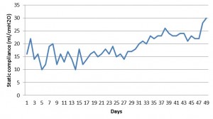
Figure 1: Static lung compliance with days
DISCUSSION
Obesity can adversely alter pulmonary function through its deleterious effect on respiratory mechanics, resistance within the respiratory system, respiratory muscle function, lung volumes, work and energy cost of breathing, control of breathing and gas exchange.[1] We reviewed the obesity related respiratory changes and summarized in Table 2.
Table 2: summary of obesity related respiratory changes
|
Change in respiratory parameter |
Description |
| Decreased compliance (chest wall >> lung) | Reduced by as much as two-thirds of the normal due to increased pulmonary blood, accumulation of fat in and around the ribs, the diaphragm and the abdomen2 |
| Decrease in FEV1 to forced vital capacity (FVC) ratio, maximal inspiratory flow rate | May be related to small airway collapse due to decreased lung volumes with increasing obesity.3,4 |
| Increase work of breathing | 70% more than normal.5 |
| Respiratory muscle dysfunction | Maximal voluntary ventilation (MVV) is reduced by 20% to 45% result from diaphragm dysfunction due to increased abdominal and visceral adipose tissue and fiber overstretching.1,5-6 |
| Ventilation perfusion inequality | Related to the degree of reduction in ERV, secondary to airway closure and alveolar collapse.1 |
| Decreased compliance (chest wall>>lung) | Reduced by as much as two-thirds of the normal due to increased pulmonary blood, accumulation of fat in and around the ribs, the diaphragm and the abdomen.2 |
| Decrease in FEV1 to forced vital capacity (FVC) ratio, maximal inspiratory flow rate | May be related to small airway collapse due to decreased lung volumes with increasing obesity.3,4 |
| Increase work of breathing | 70% more than normal.5 |
| Respiratory muscle dysfunction | Maximal voluntary ventilation (MVV) is reduced by 20% to 45% result from diaphragm dysfunction due to increased abdominal and visceral adipose tissue and fiber overstretching.1,5-6 |
| Ventilation perfusion inequality | Related to the degree of reduction in ERV, secondary to airway closure and alveolar collapse.1,5 |
Mechanical ventilation in obese patients:
We ventilated the patient for 52 days. It has been observed that due to the mechanical and inflammatory alterations observed in obesity, this subgroup of patients usually needs prolonged ventilation.7 Mechanical ventilation in this population requires specific ventilator settings. We initially ventilated the patient with SIMV+PS than switched over to PCV-VG. No data suggest that any particular mode of ventilation is superior to any other mode in patients with obesity. Marik and Varon recommend calculating the initial tidal volume according to ideal body weight (IBW) and then making adjustments on the basis of airway pressures (plateau pressure) and the results of arterial blood gas analysis.8 In order to reduce lung stress and strain, as well as minimize the risk of ventilator associated lung injury, Leme Silva P et al suggested the following ventilator strategies:
1. Stepwise recruitment maneuver before positive end-expiratory pressure application, which requires titration according to respiratory system dynamic compliance.
2. Tidal volume (VT) titration according to inspiratory capacity.9
Gizella I. Bardoczky et al studied eight morbidly obese patients during general anesthesia and controlled mechanical ventilation and observed that increasing tidal volume although caused recruitment of alveolar units, but there was no significant improvement in oxygenation.10
Weaning from ventilation was a great challenge to us. It has been suggested that the reverse Trendelenburg position at 45° can facilitate the weaning process by allowing a larger tidal volume and lower respiratory rates. El-Solh AA et al used noninvasive ventilation in immediate post-extubation period in 62 obese patients. They found significant reduction in post-extubation respiratory failure with a reduced length of ICU stay.11 We also considered noninvasive ventilation for our patient if patient had any respiratory difficulty after extubation. But fortunately, our patient did not require it.
We performed early tracheostomy anticipating need of prolonged mechanical ventilation. It is still debatable whether percutaneous tracheostomy is preferable to open surgical tracheostomy in obese patient. Ali A. ElSolh et al observed that morbid obesity is associated with increased frequency of life-threatening complications from conventional tracheostomy compare to control group.12 Percutaneous tracheostomy is also not without hazards. Various problems e.g. difficulty in identification of anatomical land marks, to obtain proper position and possibility of creating a false track may be encountered during percutaneous tracheostomy. Naresh G Mansharamani et al successfully performed percutaneous tracheostomy in 13 obese patients with the mean BMI of 45.9 ± 12.4 kg/m2.13
Our patient had also suffered from hypothyroidism which is commonly associated with obesity and is also a cause of prolonged ventilation. Datta D et al studied 140 patients receiving prolonged mechanical ventilation with failure to wean. 4% of these patients had hypothyroidism whose weaning was possible after thyroid supplements.14 We were lucky to sail through turbulent waters safely and successfully weaned off the patient after prolonged ventilation against all odds. Patience and persistent vigilance were the keys to the success.
REFERENCES
1. Koenig SM. Pulmonary complications of obesity. Am J Med Sci. 2001 Apr;321(4):249-79 [Medline]
2. Naimark A, Cherniack RM. Compliance of the respiratory system and its components in health and obesity. J Appl Physiol. 1960 May;15:377-82. [Medline]
3. Rubinstein I, Zamel N, DuBarry L, Hoffstein V. Airflow limitation in morbidly obese, nonsmoking men. Ann Intern Med. 1990 Jun 1;112(11):828-32. [Medline]
4. Lazarus R, Sparrow D, Weiss ST. Effects of obesity and fat distribution on ventilatory function: the normative aging study. Chest 1997 Apr;111(4):891-8 [Medline].
5. Ray CS, Sue DY, Bray G, Hansen JE, Wasserman K. Effects of obesity on respiratory function. Am Rev Respir Dis 1983 Sep;128(3):501-6. [Medline]
6. Sharp JT, Druz WS, Kondragunta VR. Diaphragmatic responses to body position changes in obese patients with obstructive sleep apnea, Am Rev Respir Dis 1986 Jan;133(1):32-7 [Medline]
7. Ali El-Sohl et al. Morbid obesity in medical ICU, Chest. 2001;120(6):1989-1997. [Medline]
8. Marik P, Varon J. The obese patient in the ICU. Chest 1998;113:492–498 [Medline]
9. Leme Silva P, Pelosi P, Rocco PR. Mechanical ventilation in obese patients. Minerva Anestesiol 2012 Oct;78(10):1136-45. [Medline]
10. Gizella I. Bardoczky. Large Tidal Volume Ventilation Does Not Improve Oxygenation in Morbidly Obese Patients During Anesthesia. Anesth Analg 1995;81:385-8. [Medline]
11. El-Solh AA. Noninvasive ventilation for prevention of post-extubation respiratory failure in obese patients. Eur Respir J 2006 Sep;28(3):588-95. [Medline] [Free Full Text]
12. Ali A El Solh, Wafaa Jaafar. A comparative study of the complications of surgical tracheostomy in morbidly obese critically ill patients. Critical Care 2007;11(1):R3. [Medline] [Free Full Text]
13.Mansharamani NG, Koziel H, Garland R, LoCicero J 3rd, Critchlow J, Ernst A. Safety of bedside percutaneous dilataional tracheostomy in obese patient in ICU. Chest 2000;117(5):1426-1429. [Medline]
14. Datta D, Scalise P. Hypothyroidism and failure to wean in patients receiving prolonged mechanical ventilation at a regional weaning center. Chest. 2004 Oct;126(4):1307-12. [Medline] [Free Full Text]
