
Divya Jain, MD*,Supriya Sampley, MD**, Gurpreet Kaur, MD**
*Assistant Professor, **Senior resident
Department of Anesthesiology and Intensive Care, Postgraduate Institute of Medical Education and Research, Chandigarh (India)
Correspondence: Dr Divya Jain, Assistant Professor, Department of Anesthesiology and Intensive Care, Postgraduate Institute of Medical Education and Research, Chandigarh (India); Phone: 09855900171; E-mail: jaindivya77@rediffmail.com
ABSTRACT
VACTERL association is a notable syndrome for various congenital malformations, occurring in neonates. These neonates usually present for corrective surgery of one or more malformations. We report here a rare case of VACTERL spectrum with cleft lip and palate along with hypoplastic mandible, undergoing surgery for imperforate anus under general anesthesia.
Key words: VACTERL spectrum; VACTERL association; Cleft lip; Cleft palate; Imperforate anus
Citation: Jain D, Sampley S, Kaur G. Association of difficult airway to VACTERL anomaly: An anesthetic challenge. Anaesth Pain & Intensive Care 2013;17(2):—
INTRODUCTION
The VATER/VACTERL association is a syndrome that has characteristic congenital anomalies involving vertebral malformations, anal atresia, cardiovascular anomalies, tracheoesophageal fistula, esophageal atresia, renal malformations, and dysplasias of the limbs.1-3 The estimated incidence is at ~1 in 10000 – 40000 births.4 Anesthetic management of these babies is challenging in view of multiple considerations with regards to cardiac, renal and tracheoesophageal anomalies. Airway management may be difficult anatomically and may require careful planning. We report a case of VACTERL anomaly with cleft lip and palate along with hypoplastic mandible which was operated for imperforate anus. We have discussed the anaesthetic considerations which need to be taken for the management of such cases.
CASE REPORT
A two days old neonate presented to the pediatric emergency with chief complaints of abdominal distension since 4 hrs of life and non passage of stools since birth. The neonate was a term normal, home vaginal delivery and was accepting feeds well till day 2 of life. The mother noticed abdominal distension once the baby was breast fed. The pediatric surgeon evaluated and diagnosed the neonate to have imperforate anus on abdominal x-rays and colostomy was planned.
A complete preanesthetic evaluation was done. On clinical examination the infant weighed 1.8 kg and had bilateral cleft lip and palate Grade IV along with bilateral radial hypoplasia. On chest auscultation, S1 and S2 were heard with soft systolic murmur. In respiratory system, bilateral normal vesicular breathing was auscultated. No pallor and icterus was noted and the spine appeared to be normal (Figure 1).
The patient was classified as ASA III in view of multiple anomalies. Patient was taken into operating room and baseline monitors were attached including electrocardiography (ECG), non-invasive blood pressure (NIBP), pulse oximetry and precordial stethoscope. Anticipating difficult airway in view of grade IV cleft lip and palate, a difficult airway cart was kept ready. This included mask of sizes varying from 00 to 1, Rendel Baker mask, oral airway, intubation stylets, intubation bougie of appropriate size and assorted sizes of endotracheal tubes (size 2.5, 3.0, 3.5) and Ring-Adair-Ebstein (RAE) endotracheal tubes of similar sizes; and large bore suction was kept ready. Patient already had an intravenous access in situ at the foot.
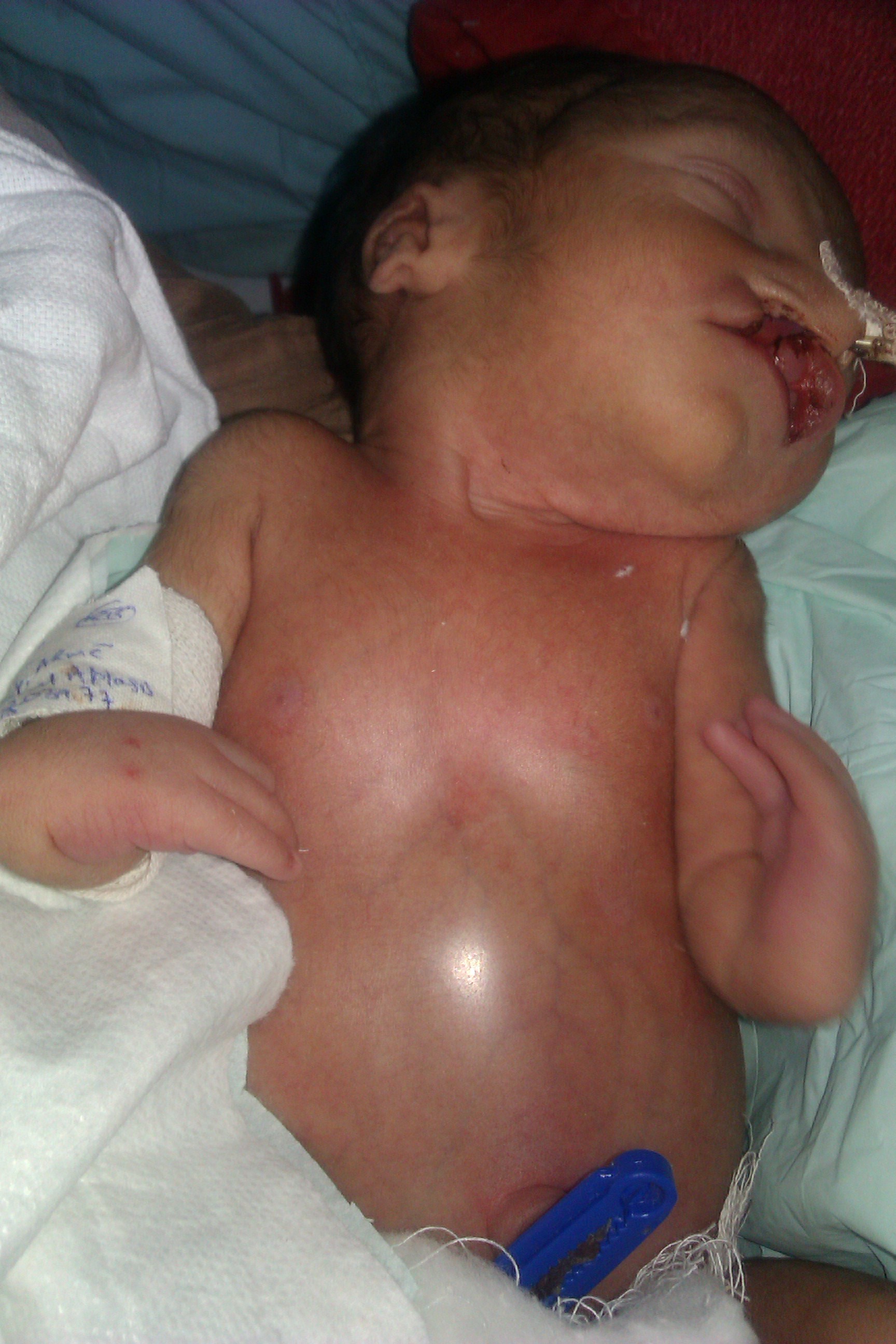
Figure 1: Picture of the newborn showing cleft lip and palate, abdominal distension and radial dysplasia
Taking into consideration the intestinal obstruction due to imperforate anus, abdominal distension and anticipated difficult airway, we planned to go for inhalational induction. After adequate preoxygenation with 100% oxygen, the patient was induced with sevoflurane, starting in concentration of 1% to 2% in 100% O2 and increased in small increments to 6%. Once the patient was induced, a check laryngoscopy was performed. Only a very small portion of posterior cords was visible where movement of air could be felt. Depth of anesthesia was increased and the trachea was intubated using 2.5 mm endotracheal tube mounted on a stylet, with an assistant employed to deliver BURP manoeuvre. Bilateral air entry was checked and found to be equal on both sides. Tube was fixed along the cleft at alae of the nose. Inj. fentanyl 2 µg/kg was given intravenously and inj. atracurium was given as a neuromuscular relaxant. Intermittent suction through the nasogastric tube helped prevent aspiration and deflated the distended abdomen. After the surgery, the patient was put on mechanical ventilation according to the weight and age parameters. The surgery went uneventfully. After the surgical procedure was over, neuromuscular blockade was reversed and the trachea extubated. Patient was shifted to pediatric intensive care unit in stable condition.
DISCUSSION
The term ‘VACTERL’ describes a group of anomalies which often occur together in newborn babies.
The association was first proposed by Quan and Smith in 1972, initially as the acronym VATER.5 Subsequently, in 1975, the VATER acronym was expanded to VACTERL which includes vertebral anomlies-70%, anal anomalies-80%, cardiac anomalies-53%, tracheo-esophageal atresia-70%, renal anomalies-53%, and limb anomalies-65%.6 To qualify as a ‘VACTERL’ child, three of the seven components mentioned above must be present. There may also be other characteristics which occur more frequently in affected children than the rest of the population; these include ear abnormalities, genital anomalies, cleft lip and/or palate, thumb anomalies, various changes in gut development and in the fetus, and the presence of a single artery in the umbilical cord. Craniofacial malformations such as choanal atresia, macrostomia, and facial hypoplasia have been associated with esophageal atresia.7 The malformations of face and esophagus may be explained by disturbances in the development of the embryo between 5th and 10th weeks of intrauterine life.8
Our patient was included into VACTERL group as the patient had anorectal malformation, bilateral radial hypoplasia and a systolic murmur. Other entities of VACTERL association were ruled out as there was no significant cardiac abnormality or any history suggestive of renal anomaly. To the best of our knowledge, no case report of any VACTERL anomaly with cleft lip and palate in anesthesia literature has been reported, owing to its extreme rare prevalence. Association with cleft lip and palate in our case was significant to be reported. A Hungarian population-based dataset of multiple malformations including orofacial clefts, have shown that 10% of the population with craniofacial clefts had a significant association with multiple congenital syndromes, out of which VACTERL is one of the likely association.
In view of multiple systemic considerations, such cases have always been very challenging to an anesthesiologist. Airway management requires appropriate planning in view of anatomical variations. Aspiration prophylaxis is warranted in such patients given their predisposition to aspiration, and the possible difficulty with airway management. This case posted for anorectal malformation was a case with trouble at both ends. Anticipated problems were difficult airway with abdominal distension, so the patient was kept nil by mouth with a nasogastric tube in situ. Positive pressure ventilation was avoided due to increased risk of regurgitation of gastric contents. The use of a short acting muscle relaxant was deferred in view of difficult mask ventilation. Check laryngoscopy under inhalational induction helped us to deal with airway. Further patients with cleft palate usually have difficulty in swallowing and recurrent episodes of aspiration followed by chest infections. In our patient the chest was clear, with no sign of respiratory infection. So the decision of extubating the child on table was made.
Other options available in difficult airway situation include various intubating LMA’s, e.g. Fastrack™ ((The Laryngeal Mask Company, Limited, San Diego, CA) and air-Q™ etc. Fibroptic bronchoscope aided intubation is another option, although in cleft palates it is not always easy.9-12 These devices are an integral part of the difficult airway trolley, but were not needed in our case as intubation was successful in single attempt with the aid of a stylet moulded into a hockey shape and the BURP manoeuvre.14 Of course, awake intubation was not an option available to us due to age of the neonate.
Postoperative problems include postextubation croup and obstructive sleep apnea. Extubating the infant or child with a difficult airway needs to be orchestrated as carefully as intubating the infant or child with a difficult airway.13 Many of the neonates and small children need postoperative ventilation for a variable period of time. Our neonate had a smooth and uneventful recovery, probably due to successful intubation in a single attempt and avoidance of excessive airway handling.
CONCLUSION
In conclusion patients with VACTREL anomaly must undergo complete preoperative assessment to rule out all the entities involved in this syndrome. The surgical intervention is usually required for the normal development of the child e.g., correction of cleft lips and palate, vertebral and anorectal correction. The airway and anesthetic management in such patients depends on the type, extent and severity of craniofacial-vertebral anomalies, associated cardiovascular problems and nature of surgery. The anesthesiologists need to formulate a well-thought of plan for securing the airway and all available airway management modalities must be kept ready.
REFERENCES
- Lawhon SM, MacEwen GD, Bunnell WP. Orthopaedic aspects of the VATER association. J Bone Jt Surg, Am.1986; 68:424–9. [PubMed]
- Muraji T, Mahour GH. Surgical problems in patients with VATER associated anomalies. J Pediatr Surg.1984; 195:550–4. [PubMed]
- Weber TR, Smith W, Grosfeld JL. Surgical experience in infants with the VATER association. J Pediatr Surg 1980; 15(6):849–54. [PubMed]
- Solomon B. VACTERL/VATER Association. Orphanet J Rare Diseases 2011; 6:56. [PubMed] [Free Full Text]
- Quan L, Smith DW. The VATER association. Vertebral defects, anal atresia, T-E fistula with esophageal atresia, radial and renal dysplasia: a spectrum of associated defects. J Pediatr 1973; 82:104–7. [PubMed]
- Khatavkar SS, Jagtap S R. Anaesthetic management of VACTERL for cataract surgery. Indian J Anaesth 2009; 53:94-7. [PubMed] [Free Full Text]
- Brereton RJ. Skeletal anomalies in oesophageal atresia. Z Kinder-chirurgie 1979; 26:258-70.
- Azmy AF, Raine P, Young DG. Orofacial clefts and oesophageal atresia. Arch dis child 1983; 58:639-41. [PubMed] [Free Full Text]
- Brain AIJ, Verghese C. LMA-Fastrack™ instruction manual. San Diego, Laryngeal Mask Company, 1998.
- Khanna P, Baidya DK, Tomar V, Agarwal A. Successful use of air-Q intubating laryngeal airway after failed rapid sequence intubation in a child with Rubinstein-Taybi syndrome. Indian J Anaesth. 2013 Mar;57(2):203-4. [PubMed] [Free Full Text]
- Jagannathan N, Wong DT. Successful tracheal intubation through an intubating laryngeal airway in pediatric patients with airway hemorrhage. J Emerg Med. 2011 Oct; 41(4):369-73. [PubMed]
- Sinha R, Chandralekha, Ray BR. Evaluation of air-Q™ intubating laryngeal airway as a conduit for tracheal intubation in infants–a pilot study. Paediatr Anaesth. 2012 Feb;22(2):156-60. [PubMed]
- Infosino A. Pediatric upper airway and congenital anomalies. Anesthesiology Clin N Am 2002; 20:747-766. [PubMed]
- Onda M, Inomata S, Satsumae T, Tanaka M. The efficacy of the “BURP” maneuver during laryngoscopy and training period necessary for residents in anesthesiology. Masui. 2012 Apr; 61(4):444-7. [PubMed]
