
M Iqbal Memon*, M Ashraf**
* Professor & HoD, **Associate Professor
Dept. of Anesthesiology & Intensive Care, Pakistan Institute of Medical Sciences, Islamabad (Pakistan).
Correspondence: Professor M Iqbal Memon,Dept. of Anesthesiology & Intensive Care, Pakistan Institute of Medical Sciences, Islamabad (Pakistan); Cell: 0333-5273054; E-mail: memoniqbal@hotmail.com
ABSTRACT
A 45 years old female with huge goiter presented for thyroidectomy. She had history of snoring and dysnea, airway evaluation revealed Mallampatti 2, IIG 3F, but temporomendibular distance (TMD) and sternomendibular distance (SMD) were impossible to measure due to grossly enlarged thyroid. Radiological examination revealed displacement of trachea towards right. After induction and depolarizer relaxation laryngoscopy was performed and Cormack Lehane 4, POGO zero was observed. With our technique of antigravity lifting, vocal cords were visualized making intubation possible with endotracheal tube on stylet.
Key words: Difficult airway; antigravity lift
Citation: Memon MI, Ashraf M. Antigravity lift technique is helpful in difficult intubation in patients with large goiters (Case report). Anaesth Pain & Intensive Care 2009;13(2):78-80
INTRODUCTION
Difficult or even failed intubation can be a major source of morbidity and mortality in clinical practice, particularly in patients with large goiters, depending upon the size of the swelling and associated compression of airways. The later constitute an aggravating factor for difficult intubation. We present a case report of a patient, who was anticipated as a case of difficult laryngoscopy and intubation. The patient presented for thyroidectomy in a camp with huge goiter and due to non-availability of fiberoptic device, we planned to manage the intubation as mentioned. We used the technique of antigravity lift technique devised by the authors in post earthquake 2005 period.
CASE REPORT
A young female of 45 years, presented for thyroidectomy with huge goiter (in euthyroid status) producing compressive symptoms, i.e. snoring and dyspnoea (Fig 1). Airway
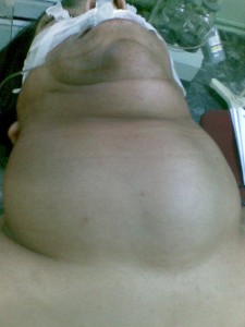
Figure 1: Picture showing the size and extent of the goiter
evaluation revealed Mallampatti II and inter-incisor gap of 3 fingers; TMD and SMD could not be measured due to enlarged thyroid. Radiological examination revealed an endothoracic goiter and displacement of trachea on right side (Fig 2) but without any compression. Deviation of trachea was defined as midline deviation of more than 1 cm.1
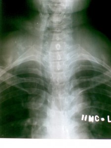
Figure 2: AP radiograph shows marked deviation of the trachea but no constriction or compression
In the operating room difficult airway trolley was arranged and patient’s NIBP, SpO2, and ECG were monitored. After pre-oxygenation for five minutes, anesthesia was induced with propofol 2.5 mg/Kg followed by suxamethonium 100mg. Patient was difficult to ventilate even after placing Gudel’s airway. On laryngoscopy, Cormack Lehane 4, POGO zero was noted. An assistant had already been explained the technique to support lateral margins of swelling and lifting against the gravity with gentle pressure (Fig 3). With this technique of antigravity lift, vocal cords were visualized and intubation was easily accomplished with 7.0 mm endotracheal tube threaded on a stylette. Correct positioning of endotracheal tube was confirmed by bilateral auscultation. Thyroidectomy was performed with patient in supine position with the head slightly extended. At the end of the procedure patient was reversed with standard drugs in practice. After return of spontaneous breathing and response to verbal command, extubation was performed. Intubation related injury to right tonsillar pillar was noted. Patient was shifted to PACU for monitoring.
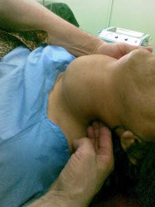
Figure 3: Technique of lifting the mass to assist in intubation
DISCUSSION
Goiter when accompanied by tracheal compression constitutes an important risk factor for difficult airway, but usually no resistance is encountered in passage of the endotracheal tube through the compressed portion of the trachea.2-5 In a comparative study of goiter patients with patients having no evidence of any risk factor, incidence found for difficult airway was 6.8% vs. 0.9%.3 In another study, patients undergoing thyroid surgeries were evaluated by IDS (1). Overall incidence was found to be 11.1 % in goiter patients.6 Whereas percentage of difficult intubation with an IDS>5 (moderate to major difficulty of intubation) was found as 5.3% and the rate of easy tracheal intubation (IDS= 0) was 36.9%. 57.8% had minor difficulty with intubation. The factors associated with goiter linked to DEI were cancerous goiter, tracheal deviation or compression, and presence of dyspnea.7 Many articles have been published suggesting that goiter, when accompanied by airway deformity, constitutes an aggravating factor for DEI.3,4,8 Intubation difficulty is commonly identified as risk factor for morbidity and mortality.9 The airway management in these cases varies according to signs, symptoms, clinical and radiological findings. Common technique for managing difficult laryngoscopy is posterior displacement of the larynx by putting backward pressure on the thyroid or cricoid cartilage (BACK maneuver). This maneuver reduces the incidence of failure to view any portion of the glottis from about 9.2% to 1.6%.10 Displacement of the larynx in three specific directions: (a) posteriorly against the cervical vertebrae (b) as far superior as possible and (c) slightly laterally to the right (BURP)11-13 and the use of a stylette or bougie have all been tried. Fiberoptic devices have revolutionized the management of difficult airway in many situations.
In cases with very large goiters, where BACK and BURP are not possible i.e. trachea is collapsed under influence of relaxant and weight of swelling, our presently reported technique consisting of antigravity raising of goiter can improve glottis exposure and ease the intubation to secure the airway. We have successfully managed three similar patients using this technique in our practice till now.
REFERENCES
- Adnet F, Borron SW, Racine SX, Clemessy JL, Fournier JL, Plaisance P, Lapandry C. The intubation Difficulty Scale (IDS): proposal and evaluation of a new score characterizing the complexity of endotracheal intubation. Anesthesiology 1997;87(6):1290-1297.
- Benumof JL. Management of the difficult adult airway. With special emphasis on awake tracheal intubation. Anesthesiology 1991 Dec;75(6):1087-110.
- Voyagis GS, Kyriakos KP. The effect of goiter on endotracheal intubation. Anesth Analg. 1997;84(3):611-2.
- Lacoste L, Gineste D, Karayon J, Montaz N, Lehuede MS, Girault M, et al. Airway complications in thyroid surgery. Ann Otol Rhinol Laryngol 1993;102:441-6
- SahaAR, Burnett C, Alfonso A, Jaffe BM. Goiters and airway problems. Am J Surg 1989;158:378-80.
- Amathieu R, Smail N, Catineau J, Poloujadoff MP, Samii K, Adnet F. Difficult intubation in thyroid surgery: myth or reality. Anesth Analg 2006;103(4):965-8.
- Bouaggad A, Nejmi SE, Bouderka MA, Abbasi O. Prediction of difficult tracheal intubation in thyroid surgery. Anesth Analg 2004;99:603-606.
- Abdel Rahim AA, Ahmed ME, Hassan A. respiratory complication after thyroidectomy and the need for tracheostomy in patients with a large goiter. Br J Surg 1999;86:88-90.
- Caplan Ra, Benumof JL, Berry Fa et al. & ASA. Practice guidelines for management of difficult airway. A report by the American Society of Anesthesiologists Task Force on Management of the Difficult Airway. Anesthesiology 1993;78:597-602.
- Wilson M, Spiegelhalter D, Robertson J, Lesser P. Predicting difficult intubation. Brit J Anaesth 1988;61:211-6
- Knill RL. Difficult laryngoscopy made easy with a “BURP”. Can J Anaesth 1993;40:279-282.
- Takahata O, Kubota M, Mamiya K, Akma Y, Nozaka T, Matsumoto H, and Ogawa H. The efficacy of the “BURP” maneuver during a difficult laryngoscopy. Anesth Analg 1997;84:419-21.
- Snider DD, Clarke D, RT and Finucane BT. The “BURP” maneuver worsens the glottic view when applied in combination with cricoid pressure. Can J Anaesth 2005;52:100-104.
