
Ajinkya Bhosle1, Vaishali Mohod1, Krishnarao Bhosle2, Afroz Quazi1
1Department of Anesthesiology, Grant Medical College and Sir J.J. Hospital.
2Department of Cardio-thoracic and Vascular Surgery, Grant Medical College and Sir J.J. Hospital.
Correspondence: Dr. Ajinkya Bhosle, 10, Trimurti, J.J. hospital, Byculla, Mumbai – 400008, (India); Phone: +91-9867707626; E-mail: ajinkya2302@gmail.com
ABSTRACT
Foreign body (FB) in tracheobronchial tree is a serious and potentially fatal condition in all age groups. Early diagnosis and removal are imperative to prevent mortality as well as complications.
A 45 year old heroine (diamorphine) addict was brought to the emergency department in a gasping state, with a history of substance abuse. The patient was intubated with Portex® cuffed endotracheal tube No. 8 and was shifted to intensive care unit for ventilation. Follow up x-ray chest revealed a metallic wire like FB in the bronchus. In the operating room, rigid bronchoscopy under general anesthesia failed to remove FB. Fibreoptic bronchoscope revealed an intubation stylet below carina. Subsequent attempts were unsuccessful so tracheostomy was done and the stylet was removed through tracheal stoma under fibreoptic visualization. The ventilation strategies used being ventilating bronchoscope, intermittent mask ventilation and intubation twice.
Keywords: Foreign body; Bronchoscopy; Tracheostomy; Difficult intubation
Citation: Bhosle A, Mohod V, Bhosle K, Quazi. An unusual foreign body in tracheobronchial tree:
A case report. Anaesth Pain & Intensive Care 2016;20(1):80-82.
INTRODUCTION
Foreign body (FB) in tracheobronchial tree is a serious and potentially fatal emergency at any age .1 In adults, FB in bronchus is comparatively a rare occurrence. If the object is large enough to cause complete or nearly complete obstruction of the main airway, asphyxia ensues and may lead to death. Sometimes patient may not have any symptoms or may present with recurrent chest infections only, without a known history of FB aspiration, resulting in a delayed diagnosis. Various types of foreign bodies have been reported such as tracheostomy cannula, dental crowns, implants, metal objects, and a variety of seeds and nuts.2,3
In medical institutions, patients undergoing oropharyngeal procedures in intensive care, operating rooms or emergency department may be at risk of complications of FB aspiration. Other contributing factors are difficult intubation, poor dentition, dentures, intoxication, sedatives, and neurological or psychiatric disorders.4
We report a rare complication of an intubation stylet being broken during intubation in a patient in emergency department with retention of the broken piece of it in the tracheobronchial tree.
CASE REPORT
A 45 year-old male was brought to emergency department by relatives with signs of difficulty in breathing and altered sensorium and a history of consumption of heroine (diamorphine). At the time of admission, patient was unconscious, and his both pupils were pin-point and non-reacting to light. On chest auscultation, bilateral basilar fine crepitations were present. Considering respiratory distress, patient was intubated by direct laryngoscopy with Portex® endotracheal tube no. 8 by resident who used metallic stylet to assist intubation.
Patient was then shifted to ICU and ventilated overnight in volume control mode. His urine examination revealed high levels of benzodiazepines and opioids. On day 2 of admission, patient was conscious, oriented and thermodynamically stable, so weaned off the ventilator. A routine follow up chest x-ray film arrived after his weaning off and it showed a FB in his right main bronchus (Stylet) with bilateral infiltrates (Figure 1).
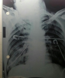
Figure 1: X-ray chest PA view showing metallic stylet
CT scan chest was ordered which suggested a metal density in trachea continuing up to his right main bronchus. It was noticeable that at the period of retention of the stylet, peak airway pressures remained within normal limits and patient did not show any signs of discomfort.
Under IV sedation and local lignocaine spray, flexible fiberoptic bronchoscopy was tried in ICU but it was unsuccessful. So patient was then taken to operating room by cardiothoracic surgeon in emergency. After adequate premedication and oxygenation, patient was induced with propofol 2 mg/kg and atracurium 0.6 mg/kg. Intermittent positive pressure ventilation on mask was then followed by ventilation through rigid bronchoscope. Rigid bronchoscopy failed to remove FB so fiberoptic bronchoscope was used and stylet was visualized just above the carina, but the attempts to retrieve it were unsuccessful so tracheostomy was done. The stylet was then removed through tracheostomy under fiberoptic bronchoscope guidance (Figure 2).
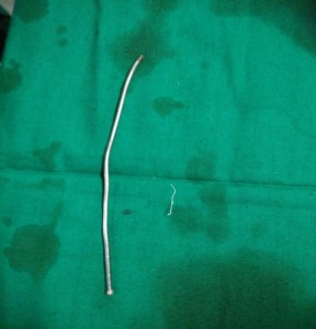
Figure 2: Broken stylet retrieved
Tracheostomy tube (TT) No. 7.5 was inserted for postoperative ventilation. The procedure required almost five hours. Anesthesia was maintained on oxygen, sevoflurane and intermittent atracurium. On postoperative day 1, chest x-ray and CT scan were repeated, which showed no evidence of FB and decreased patchy consolidation bilaterally. Ventilation was slowly weaned off and TT was removed after T-piece trial and shifted to ward on postoperative day 3.
DISCUSSION
The first case of FB removal from trachea was reported by Gustav Killian on March 30th 1897.5 In early days, FB removals from airway were mainly performed by cardiothoracic surgeons and the rigid bronchoscope was frequently utilized for this purpose. Failure to remove the FB by rigid bronchoscope was followed by thoracotomy or if necessary bronchotomy. The advent of flexible endoscopy revolutionized the care of these patients.6 Recent studies have postulated flexible bronchoscopy has higher success rate than rigid bronchoscopy.7
The common symptoms of FB aspirations are cough, fever, hemoptysis and dyspnea. Radio-opacity in chest radiography, atelectasis, emphysema and infiltration when present, should alert the attending physician to the possibility of aspirated FB.8 Three dimensional computer tomographic CT) reconstruction of aspirated FB gives exact location in tracheobronchial tree, which can be used as a guide for the endoscopist.
Intubating stylets are used for successful management of difficult airway in 78-100% patients.9 The reported complications were mucosal bleeding, sore throat etc. Broken pieces of metallic stylet resulting in partial ETT obstruction has been reported by many scholars.10-13 In our case, we observed broken stylet having migrated to right main bronchus in CT image. Thereby, we reviewed the process of intubation attempts, noticing following points: first, resident ignored signs of overuse, e.g. thinning / bending of stylet, which was possibly responsible for the breakage. Secondly, after removing the proximal part of the stylet, the resident got so busy managing the patient that he failed to pay attention to the integrity and a reduced length of the stylet by 5 cm.
The removal of tracheobronchial FB poses several considerable problems both to the endoscopist and the anesthesiologist. Several anesthesia techniques are effective for managing FB, but there is no consensus as to which technique is optimal. In this case we used IV induction and inhalational maintenance with controlled ventilation as the metal FB was too deep in right bronchus. The ventilation strategies used being intermittent mask ventilation, ventilation through rigid bronchoscope, then intubation with ET tubes twice and lastly tracheostomy. Individual in which ball-valve obstruction or bilateral FB in bronchus or chances of FB should not go inside terminal bronchus causing collapse, to avoid complete obstruction positive pressure ventilation should be avoided. Thereby, throughout the procedure we were vigilant about tracheobronchial laceration, damage, bleeding, pneumothorax and pneumomediastinum like complications.
CONCLUSION
For the segmental bronchi FB, the history should be well reviewed and a detailed investigation should be performed with immediate suspicion. Good communication between endoscopist and anesthesiologist, expertise of personnel, familiarity with equipment and clear critical plan is the key to success. Anesthesia technique has to be individualized according to requirement for every patient.
Author contribution:
AB: Conduction of the case, manuscript writing and corresponding author
VH: Supervisor, guide, manuscript editor
AQ: Conduction of Case
KB: Operating surgeon
REFERENCES
- Baharloo F, Veyckemans F, Francis C, Biettlot MP,Rodenstein DO. Tracheobronchial foreign bodies: presentation and management in children and adults. Chest. 1999 May;115(5):1357-62. [PubMed]
- Sentuk E, Sen S. An unusual case of foreign body aspiration and review of the literature. Tuberk Toraks. 2011;59(2):173-7. [PubMed] [Free full text]
- Qureshi A., Behzadi A. Foreign-body aspiration in an adult. Can J Surg. 2008 Jun; 51(3):E69-70. [PubMed] [Free full text]
- Debeljak A, Sorli J, Music E, Kecelj P. Bronchoscopic removal of foreign bodies in adults: experience with 62 patients from 1974-1998. Eur Respir J. 1999 Oct;14(4):792-95. [PubMed]
- Killian G. Meeting of the society of physicians of Freiburg. Helmboltz Zentrum Minchen.1989; 45, article 378. http://www.nickalls.org/dick/papers/thoracic/hand-bronch.pdf
- Romirez-Figueroa JL, Gochicoa-Rangel LG, Ramirez-San Juan DH, Vargas MH. Foreign body removal by flexible bronchoscopy in infants and children. Peadiatr Pulmonol. 2005 Nov;40(5):392-97. [PubMed]
- Loo CM, Hsu AA, Eng P, Ong YY. Case series of bronchoscopic removal of tracheobronchial foreign body in six adults. Ann Acad Singapore. 1998 Nov;27(6): 849-53. [PubMed]
- Apfelbaum JL, Hagberg CA, Caplan RA, Blitt CD,Connis RT, Nickinovich DG, et al. Practice guidelines for management of the difficult airway: an updated report by the American Society of Anesthesiologists Task Force on Management of the Difficult Airway. Anesthesiology. 2013 Feb;118(2):251-70. doi:10.1097/ALN.0b013e31827773b2. [PubMed] [Free full text]
- Sharma PK, Khan RM, Kaul N. An Unnoticed Broken Sheathed Metallic Stylet in an Endotracheal Tube: A case report. Sultan Qaboos Univ Med J. 2010 Apr;10(1):126–8. [PubMed] [Free full text]
- Rabb MF, Larson SM, Greger JR. An unusual cause of partial ETT obstruction. Anesthesiology. 1998;88(2):548. [PubMed] [Free full text]
- Zmyslowski WP, Kam D, Simpson GT. An unusual cause of endotracheal tube obstruction. Anesthesiology. 1989 May;70(5):883. [PubMed] [Free full text]
- Sharma A, Jain V, Mitra JK, Prabhakar H. A rare cause of endotracheal tube obstruction: a broken stylet going unnoticed–a case report. Middle East J Anaesthesiol. 2008 Feb;19(4):909–11.[PubMed]
