
Mark Howes, MBBCh, FRCA, Santhana Kannan, MD, FRCA, Miriam Namih, MBBCh
Department of Anaesthesia, City Hospital, Birmingham (United Kingdom)
Correspondence: Dr. S. Kannan, Department of Anaesthesia, City Hospital, Dudley Road, Birmingham B18 7QE. (United Kingdom); Phone: 0044 121 5074343; Fax: 0044 121 5074349; Email: kannan.gas@gmail.com
ABSTRACT
Although the use of endotracheal tube for airway control during percutaneous dilatational tracheostomy provides better airway seal, it also carries the risk of damage to cuff from the needle. This report highlights a case where the guidewire entered the trachea after traversing the cuff without associated air leak. Use of a ‘tug test’ allowed detection of the cuff impalement by the guidewire.
Key words: Airway Management; Tracheostomy; Intubation, Intratracheal
Citation: Howes M, Kannan S, Namih M. An unusual case of guidewire traversing endotracheal tube cuff during percutaneous tracheostomy. Anaesth Pain & Intensive Care 2015;19(2):156-58
INTRODUCTION
Percutaneous dilatational tracheostomy (PDT) is a common procedure in the intensive care unit. Use of in-situ endotracheal tube (ETT) for airway control has the advantage of an assured cuff seal when compared to the laryngeal mask airway. However, the additional length of the cuff and ETT below the vocal cords carries a risk of cuff puncture and impalement by needle. To minimise this risk, the ETT is withdrawn until the cuff lies immediately below the vocal cords. Although this reduces the chance of cuff puncture or ETT impalement, it does not eliminate it [1,2]. Continuous assessment of free movement of guidewire has been suggested as an indicator of its correct placement [3]. We report an instance where the guidewire traversed the cuff into the trachea, was freely mobile and visualised through the fibre optic scope. Use of a ‘tug test’ allowed detection of the traverse of the cuff by the guidewire. There have been no similar reports in the literature.
CASE REPORT
A 75 year old patient weighing around 70 kg was on mechanical ventilation for type 1 respiratory failure following pneumonia and unilateral hydro pneumothorax. PDT was planned to aid weaning. No difficulty was anticipated during pre-procedure assessment. After the pre-procedure preparation, checks, sedation and paralysis, the patient was placed in an extended neck position. The ETT was withdrawn under vision so that the cuff was positioned just below the cords. Portex Uniperc™ kit was used for PDT. Following a horizontal skin incision about 1 cm below the lower border of cricoid cartilage, blunt dissection of the subcutaneous tissues was undertaken until trachea was palpable. Trachea was punctured with the needle and air was easily aspirated. The cannula was advanced into trachea and needle removed. Although the cannula could not be visualised with fibre optic scope through the ETT, the procedure was continued since air was freely aspirated from the trachea and there was no air leak suggesting cuff rupture. The guidewire was then passed easily through the cannula and was visualised in the trachea via the fibre optic bronchoscope. The cannula was removed and dilatation of the tract commenced using the dilator over the guidewire. There was difficulty in inserting the dilator. Attempt was made to withdraw the ETT in case the tip of ETT was blocking the path of the dilator. However, there was significant resistance when ETT was pulled back. It was initially suspected that the needle and the guidewire had gone through the ETT. This would have been difficult due to the thickness of ETT. There was no audible gas leak from the mouth.
A ‘tug test’ was performed by withdrawing and pushing the ETT by about a centimetre a couple of times. This corresponded to a swing of the dilator and the guidewire in the sagittal plane. It was concluded that the dilator and guidewire were somehow attached to the ETT. The dilator and guidewire were removed. Now, there was significant audible air leak in the mouth. ETT was replaced, cuff positioned just below the vocal cords and the procedure reattempted. The second attempt was performed through the same skin incision but at a slightly caudad level and the tracheostomy tube was placed uneventfully. Correct placement was confirmed by fibre optic visualisation. Further clinical course was uneventful.

Figure 1 & 2: The probe shows the path taken by the needle and the guidewire transecting the cuff at two sites
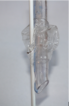
Figure 2:
DISCUSSION
Inspection of the first ETT revealed that the cuff was punctured at two sites (Figure 1 and 2). The needle, cannula and guidewire passed through the cuff, avoiding the body of the ETT itself. As the needle transfixed the cuff, there was no air leak. When the cannula went over the needle, the seal was maintained. The guidewire went into the trachea through the cannula and the transfixed cuff. This may also explain why the cannula was not visualised via the fibre optic scope.
In a study involving thirty patients, the incidence of cuff rupture and ETT impalement during PDT was reported as 6.6% each [1]. Rupture of cuff is usually immediately apparent due to significant air leak warranting corrective measures. It is rare for the cuff to be impaled without air leak.
The distance between the proximal edge of cuff to the tip of ETT ranges from 5.2 cm to 7 cm for ETT sizes ranging from 7 mm to 8.5 mm internal diameter depending upon the brand [4]. For a size 7.5 Portex ETT, this distance is typically 5.8 cm. The typical distance between vocal cords and sternal notch is around 5.3 cm when the head is resting on a pillow [5]. If no pillow is used for the head, this distance increases to 7 cm. As the first tracheal ring is about 1 cm above the sternal notch in neutral neck position, the tip of an ETT will be quite close to the site of insertion of PDT needle even if the ETT cuff is withdrawn to lie just below the vocal cords. The distance between vocal cords and sternal notch is likely to increase a bit more when neck is extended by placement of a pillow under the shoulder blades as used for PDT. This should allow the PDT needle to avoid the tip of the ETT during a puncture. However, there is considerable variation in the anatomy of patients and the distance is less in about a third of patients which increases the potential for ETT and cuff impalement. The ‘tug test’ is not mentioned in educational articles [8]although it has been quoted in one study [1]. The routine use of ‘tug test’ is important especially when fibre optic scope is not used for PDT with ETT for airway control.
This report highlights a case where the guidewire traversed the ETT cuff into the trachea without any classical signs of air leak, was freely mobile and was visualised by the fibre optic scope. The use of the ‘tug test’ helped to detect the condition.
REFERENCES
- Ambesh SP, Sinha PK, Tripathi M, Matreja P. Laryngeal mask airway vs endotracheal tube to facilitate bedside percutaneous tracheostomy in critically ill patients: a prospective comparative study. J Postgrad Med 2002;48:11-5
- Day C, Rankin N. Laceration of the cuff of an endotracheal tube during percutaneous dilatational tracheostomy. Chest 1994;105:644.
- Maddali M M, Pratap M, Fahr J, Zarroug A W. Percutaneous tracheostomy by guide wire dilating forceps technique: review of 98 patients. J Postgrad Med 2001;47:100-3
- Chong DYC, Greenland KB, Tan ST, Irwin MG, Hung CT. The clinical implication of the vocal cords–carina distance in anaesthetized Chinese adults during orotracheal intubation. Br J Anaesth 2006;97:489–95
- Wong DT1, Weng H, Lam E, Song HB, Liu J. Lengthening of the trachea during neck extension: which part of the trachea is stretched? Anesth Analg. 2008;107:989-93.
- Batuwitage B, Webber S, Glossop A. Percutaneous tracheostomy. Contin Educ Anaesth Crit Care Pain 2014;14:268 – 72
