
Sonal S. Khatavkar*, Widya S. Thatte**, Arnab Paul***
*Associate professor; **Professor & HoD; ***Resident
Department of anesthesiology, Dr. D. Y. Patil Medical Collage & Hospital, Pimpri, Pune-411018 (India)
Correspondence: Dr. Sonal S. Khatavkar, Department of anesthesiology, Dr. D. Y. Patil Medical Collage & Hospital, Pimpri, Pune-411018 (India); E-mail: drsonalkhatavkar@yahoo.co.in
ABSTRACT
Treacher Collins Syndrome (TCS) is an autosomal dominant disorder of craniofacial development. It is characterized by bilateral and symmetric abnormalities of the structures derived from the first and second branchial arch region. The patients with TCS can have difficult intubation because of retrognathia and severe facial deformity. Here we report a case of 11 months old boy with TCS with anticipated difficult airway posted for cheiloplasty under general anesthesia which was successfully managed.
Key words: Treacher Collins Syndrome; Disorders of craniofacial development; Branchial arches; Cheiloplasty; Difficult airway
Citation: Khatavkar SS, Thatte WS, Paul A. Airway management in bilateral complete cleft lip and cleft palate with features of Treacher Collins syndrome for cheiloplasty. Anaesth Pain & Intensive Care 2014;18(2):201-203
INTRODUCTION
Treacher Collins Syndrome (TCS), also known as mandibulofacial dysostosis, is an autosomal dominant disorder of craniofacial development. It involves structures derived from the first and second branchial arch region. The abnormalities are bilateral, usually symmetric, and confined to the craniofacial complex. TCS is characterized by antimongoloid slant of palpebral fissures, lower lid coloboma, retrognathia, hypoplasia of supraorbital ridges, partial absence of lower eye lashes, auricle malformation, tags of skin between the ears and angle of mouth, cleft lip and cleft palate and cryptorchidism.[1] These patients may pose difficulty in intubation because of retrognathia and severe facial deformity. So the anesthesiologist needs to be well-prepared for a difficult intubation and to maintain their airway in this often challenging patient.[2,3]
CASE REPORT
A male child of 11 months weighing 6.2 kg presented with the complaints of inability to feed and deformity of the lips and face. His was born by a full term normal delivery. He had no history of cyanotic spells at birth, repeated URTI and regurgitation. There was no history of consanguinous marriage of his parents. He was posted for cheiloplasty (plastic surgery to repair lip defects).
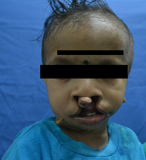
Figure 1: Child showing complete cleft lip & features of TCS
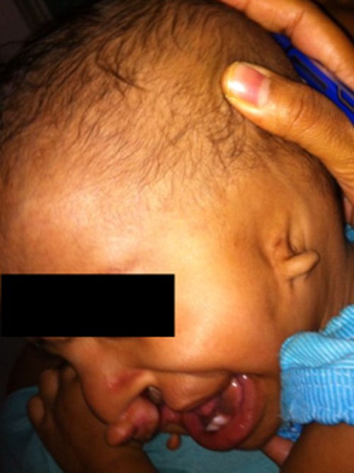
Figure 2: Absent left ear lobe and micrognathia
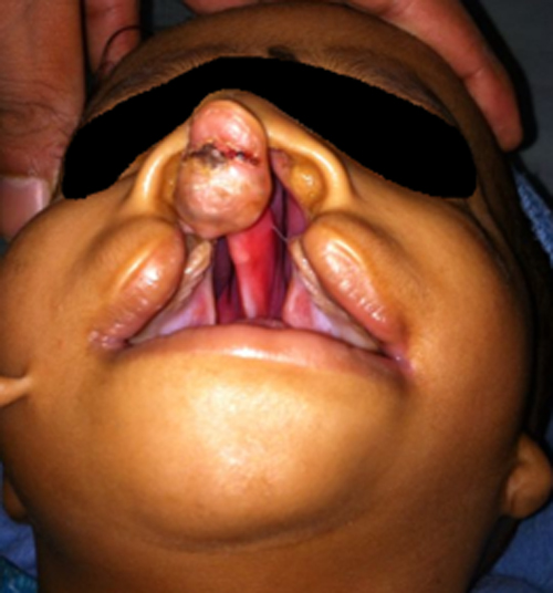
Figure 3: Showing complete bilateral cleft lip and palate
On general examination, patient was found to be afebrile with heart rate of 90 bpm and a respiratory rate of 22/min. There were complete bilateral cleft lip and palate, two uvulae, absent left ear lobe, left undescended testis, increased intercanthal distance, decreased eye lashes on lower eye lids and cutaneous tags in front of right ear and cheek. Mallampati classification could not be elicited. Neck movements were normal. Systemic examination and preoperative routine investigations were within normal limits. Hb was 14.5 gm. Blood urea, serum creatinine, serum electrolytes and liver function tests all were within normal limit. Chest x-ray (PA view) and 2D echo were normal. X-ray neck (antero-posterior/lateral views) showed a trachea displaced towards left side.
Patient was kept fasting for 4 hours. On the day of surgery an intravenous line was secured by 24G cannula in the left hand. Basic monitoring e.g. ECG, SpO2 and NIBP was done in the operating room. The child was premedicated with Inj. glycopyrrolate 0.024mg, inj. midazolam 0.12mg, inj. ondensetron 0.6mg and inj. fentanyl 12 µg IV. The patient was pre-oxygenated with 100% oxygen for 5 minutes by facemask and anesthesia was induced with sevoflurane in incremental doses starting from 2-4%. Inj propofol 14 mg was given. Bag and mask ventilation was tried and found to be adequate. We preferred inj. suxamethonium for muscle relaxation for intubation., after which IPPV was continued for 30 sec. Intubating flexible fiberoptic bronchoscope was not available with us so conventional rigid handle fiberoptic bronchoscope was used to visualize glottis opening, which was found to be anterior and shifted to left side. Miller blade No. 1 was used after failed attempts by MacIntosh blade. Larynx was still not visualized and was found to be Cormack Lehane grade IV. So backward, upward, rightward (BURP) pressure was given by a trained assistant, which converted Cormack Lehane grade to III. A cuffed RAE (south pole) tube of size 3.5 mm was tried and found to be difficult to pass. Finally trachea was intubated with uncuffed tube of size 3.5 mm. Bilateral air entry was checked and found to be equal. Throat packing was done, EtCO2 confirmed the correct placement and the tube was fixed at 9 cm. Temperature monitoring was done by rectal probe.
Anesthesia was maintained with sevoflurane 1-3% in O2 and N2O. Inj. atracurium 3.5 mg was given with subsequent top ups of 0.25 mg. To supplement anesthesia and to achieve postoperative analgesia, bilateral infraorbital block was given with 1 ml of 0.25% bupivacaine. Intraoperative vital signs remained stable. Blood loss was 30 ml and total IV fluids given were 140 ml Ringer+5% dextrose. Paracetamol suppository (80 mg) was given for postoperative analgesia.
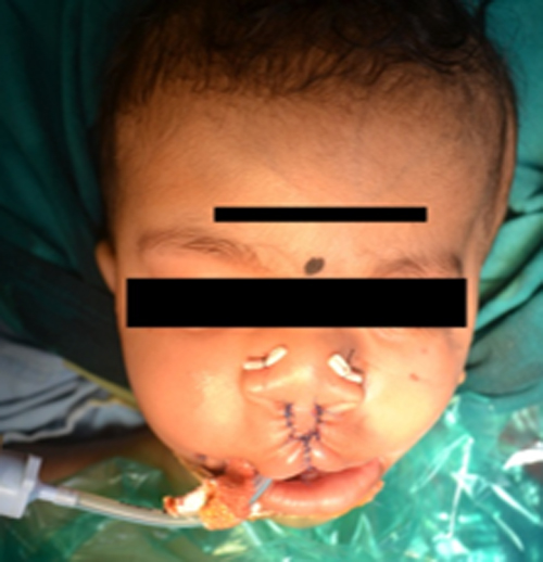
Figure 4: Intraoperative picture showing post-repair view
At the end of the surgery, muscle relaxation was reversed with neostigmine and glycopyrrolate. Throat pack removed and the child was extubated uneventfully. Postextubation vital signs were normal and shifted to PICU.

Figure 5: 3rd postop day
DISCUSSION
TCS is a rare genetic disorder characterized by craniofacial deformities. Incidence is 1 in 40,000-70,000 births.4 Cleft palate is present in upto 35% of the patients and an additional 30-40% have congenital palatopharyngeal incompetence. Abnormalities of the ears are very common and they vary from minor malformations to severe microtia and hearing loss.1 Facial clefting causes the hypoplastic appearance with possible deformities of the ear, orbital, mid face and the lower jaw areas. The clinical appearance is a result of zygoma failing to fuse with the maxilla, frontal and temporal bones.
TCS is caused by defective protein called treacle.5 More than half of the cases are thought to be due to new mutations. Mutations in the TCOF1, POLR1D gene can cause TCS. The proteins play important roles in the early development of bones and other tissues of the face. Mutations in the gene reduce the production of rRNA which may trigger the self-destruction (apoptosis) of certain cells involved in the development of facial bones and tissues.6-8
The main challenge for the anesthesiologists is maintaining airway as these patients usually have difficult tracheal intubation and regional analgesia is not the option.9 Retrognathia and relative macroglossia decrease the space available for manipulation and insertion of endotracheal tube. The associated abnormalities may be limited mouth opening, reduced extension of the head on the neck, hypoplastic mandible and limited forward movement of the hyoid.10-13 Various techniques have been described for airway management in such patients including direct laryngoscopy, intubation with a flexible fiberoptic bronchoscope, light wand, laryngeal mask airway, retrograde intubation techniques and tracheostomy.
In our patient, intubation was achieved by using a Miller blade and BURP technique. The same patient had had failed tracheal intubation three months back for cheiloplasty in a private hospital. He had classic features of Treacher Collins syndrome. We anticipated difficulty in maintaining airway as well as a difficult tracheal intubation in this child, so emergency tracheostomy tray, assorted laryngoscope blades, different types and sizes of tracheal tubes as well as other aids were kept ready.
We preferred to keep the patient breathing spontaneously without muscle relaxant but in a deep plane of anesthesia for intubation. The flexible intubating fiberoptic bronchoscope was not available with us, so we used fiberoptic bronchoscope to visualize the larynx. Intubation was successful after two attempts, with a good backward, upward and rightward pressure (BURP)[14] by a trained assistant, with the help of a Miller blade No. 1, which made visualization of glottis possible with a Cormack Lehane class III.
Flexible fiberoptic bronchoscope can be a boon for such difficult airway, but when not available then conventional approach with additional maneuvers remains the only option. Care should be taken during extubation and post operatively in view of edema of airway.
REFERENCES
- Muraika L, Heyman JS, Shevchenko Y. Fiberoptic tracheal intubation through a laryngeal mask airway in a child with Treacher Collins syndrome. Anesth Analg. 2003;97:1298-9. [PubMed]
- Ebata T, Nishiki S, Masuda A, Amaha K. Anaesthesia for Treacher Collins syndrome using a laryngeal mask airway. Can J Anaesth. 1991;38:1043-5. [PubMed]
- Bucx MJ, Grolman W, Kruisinga FH, Lindeboom JA, Van Kempen AA. The prolonged use of the laryngeal mask airway in a neonate with airway obstruction and Treacher Collins syndrome. Paediatr Anaesth. 2003;13:530-3. [PubMed]
- Andrade EC1, Júnior VS, Didoni AL, Freitas PZ, Carneiro AF, Yoshimoto FR. Treacher Collins Syndrome with choanal atresia: a case report and review of disease features. Braz J Otorhinolaryngol. 2005;71:107-10. [PubMed][Free Full Text].
- Winokur ST, Shiang R. The Treacher Collins syndrome (TCOF1) gene product, treacle, is targeted to the nucleolus by signals in its C-terminus. Hum Mol Genet. 1998;7:1947–52. [PubMed][Free Full Text]
- Bowman M1, Oldridge M, Archer C, O’Rourke A, McParland J, Brekelmans R, et al. Gross deletions in TCOF1 are a cause of Treacher-Collins-Franceschetti syndrome. Eur J Hum Genet. 2012;20:769-77. [PubMed][Free Full Text]
- So RB, Gonzales B, Henning D, Dixon J, Dixon MJ, Valdez BC. Another face of the Treacher Collins syndrome (TCOF1) gene: identification of additional exons. Gene. 2004;328:49–57. [PubMed]
- Dixon J, Ellis I, Bottani A, Temple K, Dixon MJ. Identification of mutations in TCOF1: use of molecular analysis in the pre- and postnatal diagnosis of Treacher Collins syndrome. Am J Med Genet A 2004;127A:244–8. [PubMed]
- Tsaur GR, Liaw WJ, Wong CS, Yu FK, Hwang JJ, Chen SG, Ho ST. [Treacher-Collin’s syndrome and translaryngeal guided intubation–case report]. Acta Anaesthesiol Sin 1994;32:223-228. [PubMed]
- Bellhouse CP, Dore C. Criteria for estimating likelihood of difficulty of endotracheal intubation with Macintosh laryngoscope. Anesth Intensive Care 1988;16:329-37. [PubMed]
- Charters P. Analysis of mathematical model for osseous factors in difficult intubation. Can J Anaesth 1994;41:594-602. [PubMed]
- Cormack RS, Lehane J.Difficult tracheal intubation in obstetrics. Anesthesia 1984;39:1105-11. [PubMed]
- Mallampati SR, Gatt SP, Gugino LD, Desai SP, Waraksa B, Freiberger D, et al. A clinical sign to predict difficult tracheal intubation: a prospective study. Can Anaesth Soc J 1985;32:429-34. [PubMed]
- Levitan RM1, Kinkle WC, Levin WJ, Everett WW. Laryngeal view during laryngoscopy: a randomized trial comparing cricoid pressure, backward-upward-rightward pressure, and bimanual laryngoscopy. Ann Emerg Med 2006;47:548-55. [PubMed]
